| کد مقاله | کد نشریه | سال انتشار | مقاله انگلیسی | نسخه تمام متن |
|---|---|---|---|---|
| 11014819 | 1790012 | 2018 | 12 صفحه PDF | دانلود رایگان |
عنوان انگلیسی مقاله ISI
Developing 3D microscopy with CLARITY on human brain tissue: Towards a tool for informing and validating MRI-based histology
دانلود مقاله + سفارش ترجمه
دانلود مقاله ISI انگلیسی
رایگان برای ایرانیان
موضوعات مرتبط
علوم زیستی و بیوفناوری
علم عصب شناسی
علوم اعصاب شناختی
پیش نمایش صفحه اول مقاله

چکیده انگلیسی
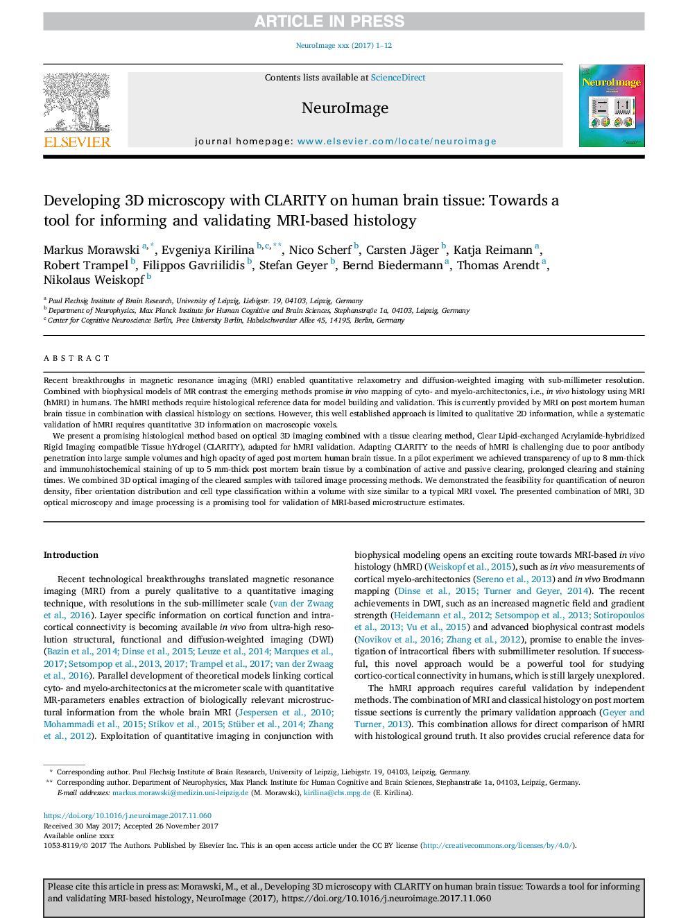
We present a promising histological method based on optical 3D imaging combined with a tissue clearing method, Clear Lipid-exchanged Acrylamide-hybridized Rigid Imaging compatible Tissue hYdrogel (CLARITY), adapted for hMRI validation. Adapting CLARITY to the needs of hMRI is challenging due to poor antibody penetration into large sample volumes and high opacity of aged post mortem human brain tissue. In a pilot experiment we achieved transparency of up to 8Â mm-thick and immunohistochemical staining of up to 5Â mm-thick post mortem brain tissue by a combination of active and passive clearing, prolonged clearing and staining times. We combined 3D optical imaging of the cleared samples with tailored image processing methods. We demonstrated the feasibility for quantification of neuron density, fiber orientation distribution and cell type classification within a volume with size similar to a typical MRI voxel. The presented combination of MRI, 3D optical microscopy and image processing is a promising tool for validation of MRI-based microstructure estimates.
ناشر
Database: Elsevier - ScienceDirect (ساینس دایرکت)
Journal: NeuroImage - Volume 182, 15 November 2018, Pages 417-428
Journal: NeuroImage - Volume 182, 15 November 2018, Pages 417-428
نویسندگان
Markus Morawski, Evgeniya Kirilina, Nico Scherf, Carsten Jäger, Katja Reimann, Robert Trampel, Filippos Gavriilidis, Stefan Geyer, Bernd Biedermann, Thomas Arendt, Nikolaus Weiskopf,