| کد مقاله | کد نشریه | سال انتشار | مقاله انگلیسی | نسخه تمام متن |
|---|---|---|---|---|
| 1231629 | 1495219 | 2015 | 13 صفحه PDF | دانلود رایگان |

• DNA- and BSA-binding studies of two water soluble copper(II) complexes.
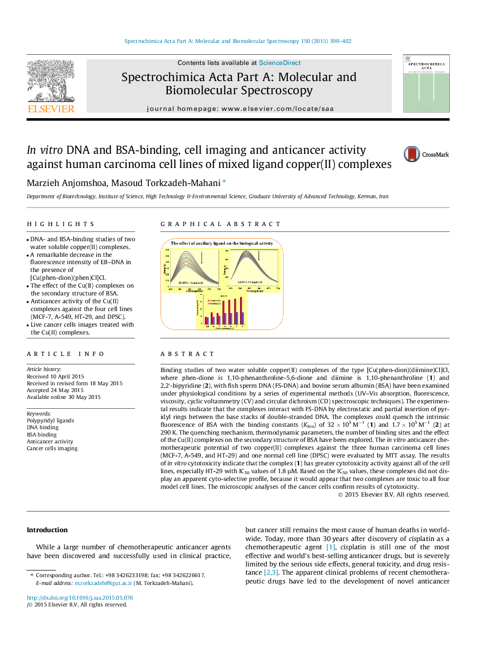
• A remarkable decrease in the fluorescence intensity of EB–DNA in the presence of [Cu(phen-dion)(phen)Cl]Cl.
• The effect of the Cu(II) complexes on the secondary structure of BSA.
• Anticancer activity of the Cu(II) complexes against the four cell lines (MCF-7, A-549, HT-29, and DPSC).
• Live cancer cells images treated with the Cu(II) complexes.
Binding studies of two water soluble copper(II) complexes of the type [Cu(phen-dion)(diimine)Cl]Cl, where phen-dione is 1,10-phenanthroline-5,6-dione and diimine is 1,10-phenanthroline (1) and 2,2′-bipyridine (2), with fish sperm DNA (FS-DNA) and bovine serum albumin (BSA) have been examined under physiological conditions by a series of experimental methods (UV–Vis absorption, fluorescence, viscosity, cyclic voltammetry (CV) and circular dichroism (CD) spectroscopic techniques). The experimental results indicate that the complexes interact with FS-DNA by electrostatic and partial insertion of pyridyl rings between the base stacks of double-stranded DNA. The complexes could quench the intrinsic fluorescence of BSA with the binding constants (Kbin) of 32 × 105 M−1 (1) and 1.7 × 10⁵ M−1 (2) at 290 K. The quenching mechanism, thermodynamic parameters, the number of binding sites and the effect of the Cu(II) complexes on the secondary structure of BSA have been explored. The in vitro anticancer chemotherapeutic potential of two copper(II) complexes against the three human carcinoma cell lines (MCF-7, A-549, and HT-29) and one normal cell line (DPSC) were evaluated by MTT assay. The results of in vitro cytotoxicity indicate that the complex (1) has greater cytotoxicity activity against all of the cell lines, especially HT-29 with IC50 values of 1.8 μM. Based on the IC50 values, these complexes did not display an apparent cyto-selective profile, because it would appear that two complexes are toxic to all four model cell lines. The microscopic analyses of the cancer cells confirm results of cytotoxicity.
Figure optionsDownload as PowerPoint slide
Journal: Spectrochimica Acta Part A: Molecular and Biomolecular Spectroscopy - Volume 150, 5 November 2015, Pages 390–402