| کد مقاله | کد نشریه | سال انتشار | مقاله انگلیسی | نسخه تمام متن |
|---|---|---|---|---|
| 2760482 | 1150174 | 2012 | 6 صفحه PDF | دانلود رایگان |

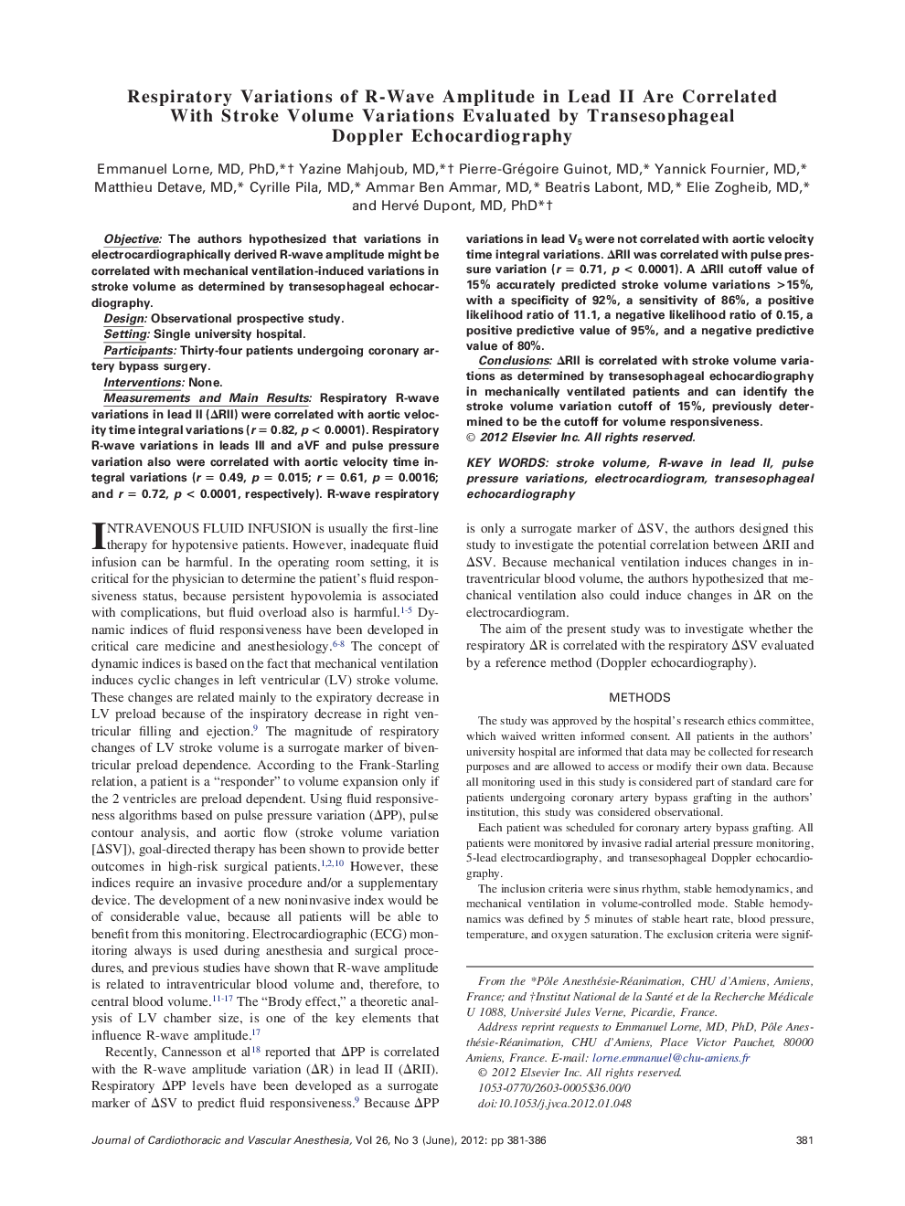
ObjectiveThe authors hypothesized that variations in electrocardiographically derived R-wave amplitude might be correlated with mechanical ventilation-induced variations in stroke volume as determined by transesophageal echocardiography.DesignObservational prospective study.SettingSingle university hospital.ParticipantsThirty-four patients undergoing coronary artery bypass surgery.InterventionsNone.Measurements and Main ResultsRespiratory R-wave variations in lead II (ΔRII) were correlated with aortic velocity time integral variations (r = 0.82, p < 0.0001). Respiratory R-wave variations in leads III and aVF and pulse pressure variation also were correlated with aortic velocity time integral variations (r = 0.49, p = 0.015; r = 0.61, p = 0.0016; and r = 0.72, p < 0.0001, respectively). R-wave respiratory variations in lead V5 were not correlated with aortic velocity time integral variations. ΔRII was correlated with pulse pressure variation (r = 0.71, p < 0.0001). A ΔRII cutoff value of 15% accurately predicted stroke volume variations >15%, with a specificity of 92%, a sensitivity of 86%, a positive likelihood ratio of 11.1, a negative likelihood ratio of 0.15, a positive predictive value of 95%, and a negative predictive value of 80%.ConclusionsΔRII is correlated with stroke volume variations as determined by transesophageal echocardiography in mechanically ventilated patients and can identify the stroke volume variation cutoff of 15%, previously determined to be the cutoff for volume responsiveness.
Journal: Journal of Cardiothoracic and Vascular Anesthesia - Volume 26, Issue 3, June 2012, Pages 381–386