| کد مقاله | کد نشریه | سال انتشار | مقاله انگلیسی | نسخه تمام متن |
|---|---|---|---|---|
| 2947200 | 1577207 | 2012 | 10 صفحه PDF | دانلود رایگان |

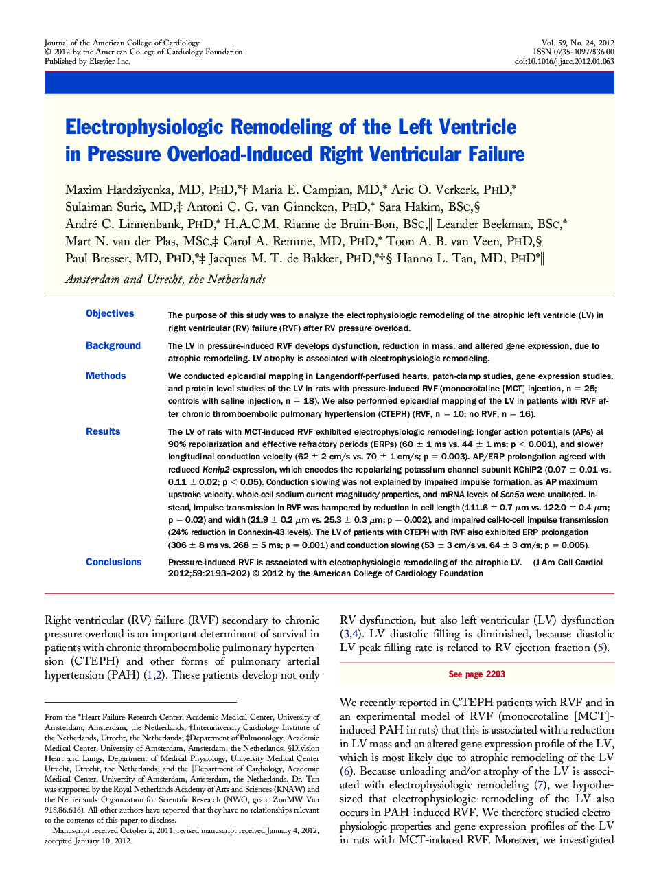
ObjectivesThe purpose of this study was to analyze the electrophysiologic remodeling of the atrophic left ventricle (LV) in right ventricular (RV) failure (RVF) after RV pressure overload.BackgroundThe LV in pressure-induced RVF develops dysfunction, reduction in mass, and altered gene expression, due to atrophic remodeling. LV atrophy is associated with electrophysiologic remodeling.MethodsWe conducted epicardial mapping in Langendorff-perfused hearts, patch-clamp studies, gene expression studies, and protein level studies of the LV in rats with pressure-induced RVF (monocrotaline [MCT] injection, n = 25; controls with saline injection, n = 18). We also performed epicardial mapping of the LV in patients with RVF after chronic thromboembolic pulmonary hypertension (CTEPH) (RVF, n = 10; no RVF, n = 16).ResultsThe LV of rats with MCT-induced RVF exhibited electrophysiologic remodeling: longer action potentials (APs) at 90% repolarization and effective refractory periods (ERPs) (60 ± 1 ms vs. 44 ± 1 ms; p < 0.001), and slower longitudinal conduction velocity (62 ± 2 cm/s vs. 70 ± 1 cm/s; p = 0.003). AP/ERP prolongation agreed with reduced Kcnip2 expression, which encodes the repolarizing potassium channel subunit KChIP2 (0.07 ± 0.01 vs. 0.11 ± 0.02; p < 0.05). Conduction slowing was not explained by impaired impulse formation, as AP maximum upstroke velocity, whole-cell sodium current magnitude/properties, and mRNA levels of Scn5a were unaltered. Instead, impulse transmission in RVF was hampered by reduction in cell length (111.6 ± 0.7 μm vs. 122.0 ± 0.4 μm; p = 0.02) and width (21.9 ± 0.2 μm vs. 25.3 ± 0.3 μm; p = 0.002), and impaired cell-to-cell impulse transmission (24% reduction in Connexin-43 levels). The LV of patients with CTEPH with RVF also exhibited ERP prolongation (306 ± 8 ms vs. 268 ± 5 ms; p = 0.001) and conduction slowing (53 ± 3 cm/s vs. 64 ± 3 cm/s; p = 0.005).ConclusionsPressure-induced RVF is associated with electrophysiologic remodeling of the atrophic LV.
Journal: Journal of the American College of Cardiology - Volume 59, Issue 24, 12 June 2012, Pages 2193–2202