| کد مقاله | کد نشریه | سال انتشار | مقاله انگلیسی | نسخه تمام متن |
|---|---|---|---|---|
| 2950107 | 1577259 | 2011 | 11 صفحه PDF | دانلود رایگان |

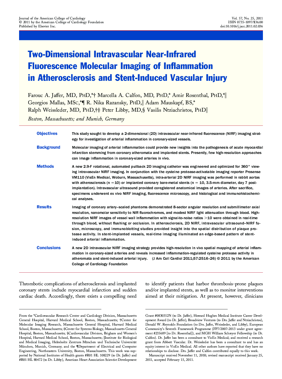
ObjectivesThis study sought to develop a 2-dimensional (2D) intravascular near-infrared fluorescence (NIRF) imaging strategy for investigation of arterial inflammation in coronary-sized vessels.BackgroundMolecular imaging of arterial inflammation could provide new insights into the pathogenesis of acute myocardial infarction stemming from coronary atheromata and implanted stents. Presently, few high-resolution approaches can image inflammation in coronary-sized arteries in vivo.MethodsA new 2.9-F rotational, automated pullback 2D imaging catheter was engineered and optimized for 360° viewing intravascular NIRF imaging. In conjunction with the cysteine protease-activatable imaging reporter Prosense VM110 (VisEn Medical, Woburn, Massachusetts), intra-arterial 2D NIRF imaging was performed in rabbit aortas with atherosclerosis (n =10) or implanted coronary bare-metal stents (n = 10, 3.5-mm diameter, day 7 post-implantation). Intravascular ultrasound provided coregistered anatomical images of arteries. After sacrifice, specimens underwent ex vivo NIRF imaging, fluorescence microscopy, and histological and immunohistochemical analyses.ResultsImaging of coronary artery–scaled phantoms demonstrated 8-sector angular resolution and submillimeter axial resolution, nanomolar sensitivity to NIR fluorochromes, and modest NIRF light attenuation through blood. High-resolution NIRF images of vessel wall inflammation with signal-to-noise ratios >10 were obtained in real-time through blood, without flushing or occlusion. In atherosclerosis, 2D NIRF, intravascular ultrasound–NIRF fusion, microscopy, and immunoblotting studies provided insight into the spatial distribution of plaque protease activity. In stent-implanted vessels, real-time imaging illuminated an edge-based pattern of stent-induced arterial inflammation.ConclusionsA new 2D intravascular NIRF imaging strategy provides high-resolution in vivo spatial mapping of arterial inflammation in coronary-sized arteries and reveals increased inflammation-regulated cysteine protease activity in atheromata and stent-induced arterial injury.
Journal: Journal of the American College of Cardiology - Volume 57, Issue 25, 21 June 2011, Pages 2516–2526