| کد مقاله | کد نشریه | سال انتشار | مقاله انگلیسی | نسخه تمام متن |
|---|---|---|---|---|
| 3119015 | 1582804 | 2006 | 5 صفحه PDF | دانلود رایگان |
عنوان انگلیسی مقاله ISI
Three-dimensional spiral computed tomographic imaging: A new approach to the diagnosis and treatment planning of impacted teeth
دانلود مقاله + سفارش ترجمه
دانلود مقاله ISI انگلیسی
رایگان برای ایرانیان
موضوعات مرتبط
علوم پزشکی و سلامت
پزشکی و دندانپزشکی
دندانپزشکی، جراحی دهان و پزشکی
پیش نمایش صفحه اول مقاله

چکیده انگلیسی
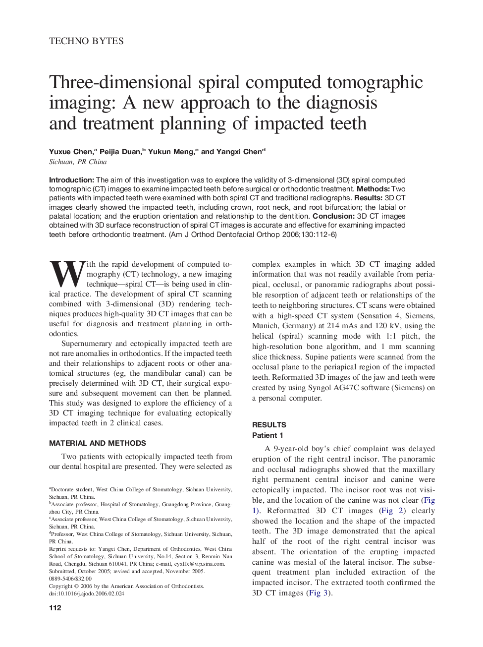
Introduction: The aim of this investigation was to explore the validity of 3-dimensional (3D) spiral computed tomographic (CT) images to examine impacted teeth before surgical or orthodontic treatment. Methods: Two patients with impacted teeth were examined with both spiral CT and traditional radiographs. Results: 3D CT images clearly showed the impacted teeth, including crown, root neck, and root bifurcation; the labial or palatal location; and the eruption orientation and relationship to the dentition. Conclusion: 3D CT images obtained with 3D surface reconstruction of spiral CT images is accurate and effective for examining impacted teeth before orthodontic treatment.
ناشر
Database: Elsevier - ScienceDirect (ساینس دایرکت)
Journal: American Journal of Orthodontics and Dentofacial Orthopedics - Volume 130, Issue 1, July 2006, Pages 112–116
Journal: American Journal of Orthodontics and Dentofacial Orthopedics - Volume 130, Issue 1, July 2006, Pages 112–116
نویسندگان
Yuxue Chen, Peijia Duan, Yukun Meng, Yangxi Chen,