| کد مقاله | کد نشریه | سال انتشار | مقاله انگلیسی | نسخه تمام متن |
|---|---|---|---|---|
| 3153524 | 1198034 | 2011 | 8 صفحه PDF | دانلود رایگان |

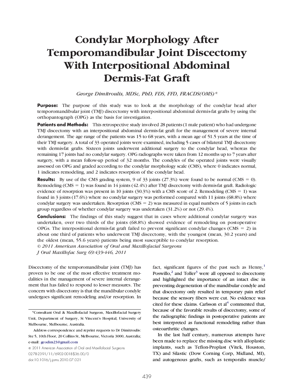
PurposeThe purpose of this study was to look at the morphology of the condylar head after temporomandibular joint (TMJ) discectomy with interpositional abdominal dermis-fat grafts by using the orthopantograph (OPG) as the basis for investigation.Patients and MethodsThis retrospective study involved 28 patients (1 male patient) who had undergone TMJ discectomy with an interpositional abdominal dermis-fat graft for the management of severe internal derangement. The age range of the patients was 15 to 68 years, with a mean age of 51.5 years at the time of their TMJ surgery. A total of 33 operated joints were examined, including 5 cases of bilateral TMJ discectomy with dermis-fat grafts. Sixteen joints underwent additional surgery to the condylar head, whereas the remaining 17 joints had no condylar surgery. OPG radiographs were taken from 12 months up to 7 years after surgery, with a mean follow-up period of 32 months. The condyles of the operated joints were visually assessed on OPG and graded according to the condylar morphology scale (CMS), where 0 indicates normal, 1 indicates remodeling, and 2 indicates resorption of the condylar head.ResultsBy use of the CMS grading system, 9 of 33 joints (27.3%) were found to be normal (CMS = 0). Remodeling (CMS = 1) was found in 14 joints (42.4%) after TMJ discectomy with dermis-fat graft. Radiologic evidence of resorption was present in 10 joints (30.3%) with a CMS score of 2. Remodeling (CMS = 1) was found in 3 joints (17.6%) where no condylar surgery was performed compared with 11 joints (68.8%) where condylar surgery was undertaken. Resorption (CMS = 2) was measured in equal numbers of 5 joints in each group regardless of whether condylar surgery was undertaken (31.2%) or not (29.4%).ConclusionsThe findings of this study suggest that in cases where additional condylar surgery was undertaken, over two thirds of the joints (68.8%) showed evidence of remodeling on postoperative OPGs. The interpositional dermis-fat graft failed to prevent significant condylar changes (CMS = 2) in about one third of patients who underwent TMJ discectomy, with the youngest (mean, 30.2 years) and the oldest (mean, 55.6 years) patients being most susceptible to condylar resorption.
Journal: Journal of Oral and Maxillofacial Surgery - Volume 69, Issue 2, February 2011, Pages 439–446