| کد مقاله | کد نشریه | سال انتشار | مقاله انگلیسی | نسخه تمام متن |
|---|---|---|---|---|
| 3174792 | 1200088 | 2006 | 15 صفحه PDF | دانلود رایگان |
عنوان انگلیسی مقاله ISI
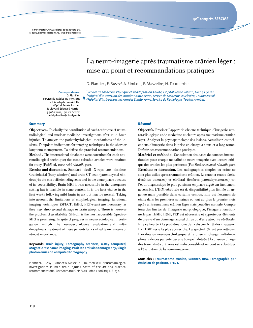
La neuro-imagerie après traumatisme crânien léger : mise au point et recommandations pratiques
دانلود مقاله + سفارش ترجمه
دانلود مقاله ISI انگلیسی
رایگان برای ایرانیان
کلمات کلیدی
IRMbrain injury - آسیب مغزیTraumatisme crânien - آسیب مغزی آسیب دیدهScanner - اسکنرTomography scanners - اسکنرهای توموگرافیX-ray computed - اشعه ایکس محاسبه شده استSPECT - برشنگاری رایانهای تک فوتونی، مقطع نگاری رایانهای تک فوتونی، توموگرافی رایانهای تک فوتونی، اسپکتMagnetic resonance imaging - تصویربرداری رزونانس مغناطیسیsingle photon emission computed tomography - توموگرافی کامپیوتری انتشار اتمی فوتونPositron emission tomography - توموگرافی گسیل پوزیترون
موضوعات مرتبط
علوم پزشکی و سلامت
پزشکی و دندانپزشکی
دندانپزشکی، جراحی دهان و پزشکی
پیش نمایش صفحه اول مقاله

چکیده انگلیسی
Standard skull X-rays are obsolete. Craniofacial (bony windows) and brain CT-scan (parenchymal windows) is the most efficient diagnosis tool in the acute phase because of its accessibility. Brain MRI is less accessible in the emergency setting but is feasible in some centers. It is the best choice in the first weeks following mild brain injury but may be normal. Taking into account the limitations of morphological imaging, functional imaging techniques (SPECT, fMRI, PET-scan) are necessary as they may show axonal damage or brain atrophy. There is however the problem of availability. SPECT is the most accessible. Spectro-MRI is promising. In spite of progress in neuroradiological investigation methods, the neuropsychological evaluation and multi-disciplinary treatment of these patients by a skilled team remains of utmost importance.
ناشر
Database: Elsevier - ScienceDirect (ساینس دایرکت)
Journal: Revue de Stomatologie et de Chirurgie Maxillo-faciale - Volume 107, Issue 4, September 2006, Pages 218-232
Journal: Revue de Stomatologie et de Chirurgie Maxillo-faciale - Volume 107, Issue 4, September 2006, Pages 218-232
نویسندگان
D. Plantier, E. Bussy, A. Rimbot, P. Maszelin, H. Tournebise,