| کد مقاله | کد نشریه | سال انتشار | مقاله انگلیسی | نسخه تمام متن |
|---|---|---|---|---|
| 4045810 | 1603611 | 2006 | 5 صفحه PDF | دانلود رایگان |
عنوان انگلیسی مقاله ISI
Arthroscopic Technique of Interposition Arthroplasty of the Glenohumeral Joint
دانلود مقاله + سفارش ترجمه
دانلود مقاله ISI انگلیسی
رایگان برای ایرانیان
کلمات کلیدی
موضوعات مرتبط
علوم پزشکی و سلامت
پزشکی و دندانپزشکی
ارتوپدی، پزشکی ورزشی و توانبخشی
پیش نمایش صفحه اول مقاله

چکیده انگلیسی
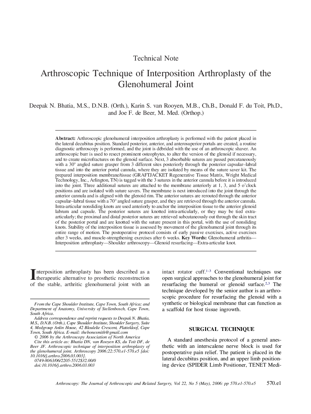
Arthroscopic glenohumeral interposition arthroplasty is performed with the patient placed in the lateral decubitus position. Standard posterior, anterior, and anterosuperior portals are created, a routine diagnostic arthroscopy is performed, and the joint is débrided with the use of an arthroscopic shaver. An arthroscopic burr is used to resect prominent osteophytes, to alter the version of the glenoid if necessary, and to create microfractures on the glenoid surface. Next, 3 absorbable sutures are passed percutaneously with a 30° angled suture grasper from 3 different sites posteriorly through the posterior capsular-labral tissue and into the anterior portal cannula, where they are isolated by means of the suture saver kit. The prepared interposition membrane/tissue (GRAFTJACKET Regenerative Tissue Matrix, Wright Medical Technology, Inc., Arlington, TN) is tagged with the 3 sutures in the anterior cannula before it is introduced into the joint. Three additional sutures are attached to the membrane anteriorly at 1, 3, and 5 o'clock positions and are isolated with suture savers. The membrane is next introduced into the joint through the anterior cannula and is aligned with the glenoid rim. The anterior sutures are rerouted through the anterior capsular-labral tissue with a 70° angled suture grasper, and they are retrieved through the anterior cannula. Intra-articular nonsliding knots are used anteriorly to anchor the interposition tissue to the anterior glenoid labrum and capsule. The posterior sutures are knotted intra-articularly, or they may be tied extra-articularly; the proximal and distal posterior sutures are retrieved subcutaneously out through the skin tract of the posterior portal and are knotted with the suture present in this portal, with the use of nonsliding knots. Stability of the interposition tissue is assessed by movement of the glenohumeral joint through its entire range of motion. The postoperative protocol consists of early passive exercises, active exercises after 3 weeks, and muscle-strengthening exercises after 6 weeks.
ناشر
Database: Elsevier - ScienceDirect (ساینس دایرکت)
Journal: Arthroscopy: The Journal of Arthroscopic & Related Surgery - Volume 22, Issue 5, May 2006, Pages 570.e1-570.e5
Journal: Arthroscopy: The Journal of Arthroscopic & Related Surgery - Volume 22, Issue 5, May 2006, Pages 570.e1-570.e5
نویسندگان
Deepak N. M.S., D.N.B. (Orth.), Karin S. M.B., Ch.B., Donald F. Ph.D., Joe F. M. Med. (Orthop.),