| کد مقاله | کد نشریه | سال انتشار | مقاله انگلیسی | نسخه تمام متن |
|---|---|---|---|---|
| 4047440 | 1603591 | 2007 | 5 صفحه PDF | دانلود رایگان |
عنوان انگلیسی مقاله ISI
Arthroscopic Anatomy and Surgical Techniques for Peritrochanteric Space Disorders in the Hip
دانلود مقاله + سفارش ترجمه
دانلود مقاله ISI انگلیسی
رایگان برای ایرانیان
موضوعات مرتبط
علوم پزشکی و سلامت
پزشکی و دندانپزشکی
ارتوپدی، پزشکی ورزشی و توانبخشی
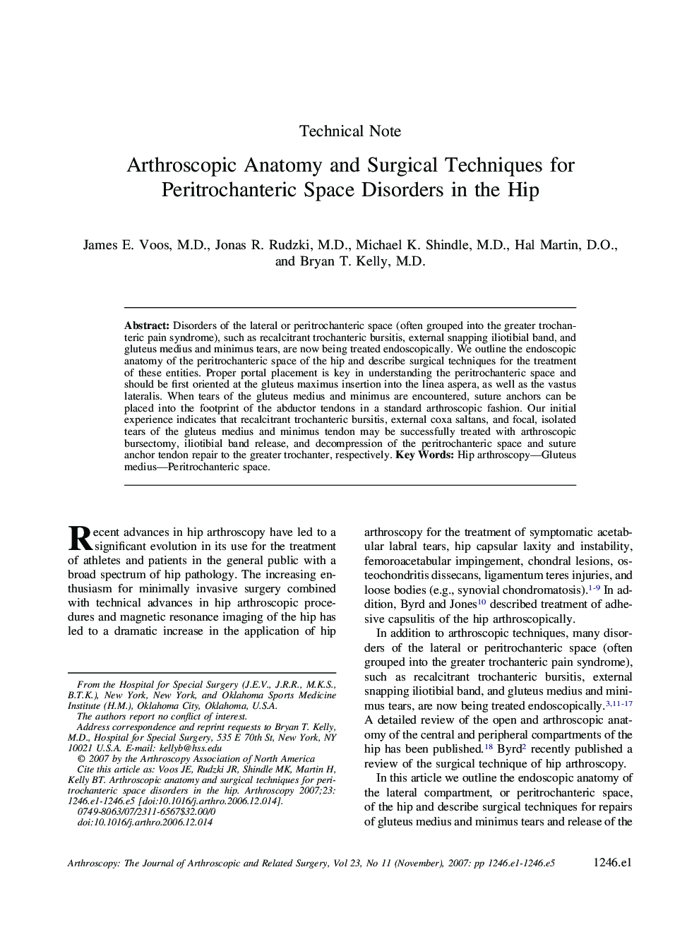
پیش نمایش صفحه اول مقاله

چکیده انگلیسی
Disorders of the lateral or peritrochanteric space (often grouped into the greater trochanteric pain syndrome), such as recalcitrant trochanteric bursitis, external snapping iliotibial band, and gluteus medius and minimus tears, are now being treated endoscopically. We outline the endoscopic anatomy of the peritrochanteric space of the hip and describe surgical techniques for the treatment of these entities. Proper portal placement is key in understanding the peritrochanteric space and should be first oriented at the gluteus maximus insertion into the linea aspera, as well as the vastus lateralis. When tears of the gluteus medius and minimus are encountered, suture anchors can be placed into the footprint of the abductor tendons in a standard arthroscopic fashion. Our initial experience indicates that recalcitrant trochanteric bursitis, external coxa saltans, and focal, isolated tears of the gluteus medius and minimus tendon may be successfully treated with arthroscopic bursectomy, iliotibial band release, and decompression of the peritrochanteric space and suture anchor tendon repair to the greater trochanter, respectively.
ناشر
Database: Elsevier - ScienceDirect (ساینس دایرکت)
Journal: Arthroscopy: The Journal of Arthroscopic & Related Surgery - Volume 23, Issue 11, November 2007, Pages 1246.e1-1246.e5
Journal: Arthroscopy: The Journal of Arthroscopic & Related Surgery - Volume 23, Issue 11, November 2007, Pages 1246.e1-1246.e5
نویسندگان
James E. M.D., Jonas R. M.D., Michael K. M.D., Hal D.O., Bryan T. M.D.,