| کد مقاله | کد نشریه | سال انتشار | مقاله انگلیسی | نسخه تمام متن |
|---|---|---|---|---|
| 4076278 | 1267081 | 2007 | 6 صفحه PDF | دانلود رایگان |
عنوان انگلیسی مقاله ISI
Computed tomography analysis of the coracoid process and anatomic structures of the shoulder after arthroscopic coracoid decompression: A cadaveric study
دانلود مقاله + سفارش ترجمه
دانلود مقاله ISI انگلیسی
رایگان برای ایرانیان
موضوعات مرتبط
علوم پزشکی و سلامت
پزشکی و دندانپزشکی
ارتوپدی، پزشکی ورزشی و توانبخشی
پیش نمایش صفحه اول مقاله

چکیده انگلیسی
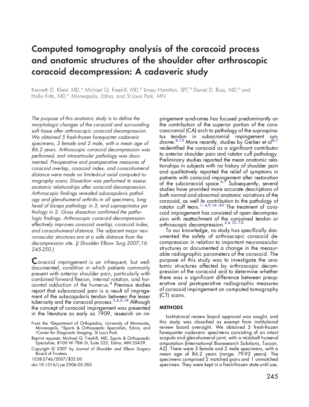
The purpose of this anatomic study is to define the morphologic changes of the coracoid and surrounding soft tissue after arthroscopic coracoid decompression. We obtained 5 fresh-frozen forequarter cadaveric specimens, 3 female and 2 male, with a mean age of 86.2 years. Arthroscopic coracoid decompression was performed, and intraarticular pathology was documented. Preoperative and postoperative measures of coracoid overlap, coracoid index, and coracohumeral distance were made on limited-cut axial computed tomography scans. Dissection was performed to assess anatomic relationships after coracoid decompression. Arthroscopic findings revealed subscapularis pathology and glenohumeral arthritis in all specimens, long head of biceps pathology in 3, and supraspinatus pathology in 2. Gross dissection confirmed the pathologic findings. Arthroscopic coracoid decompression effectively improves coracoid overlap, coracoid index, and coracohumeral distance. The adjacent major neurovascular structures are at a safe distance from the decompression site.
ناشر
Database: Elsevier - ScienceDirect (ساینس دایرکت)
Journal: Journal of Shoulder and Elbow Surgery - Volume 16, Issue 2, MarchâApril 2007, Pages 245-250
Journal: Journal of Shoulder and Elbow Surgery - Volume 16, Issue 2, MarchâApril 2007, Pages 245-250
نویسندگان
Kenneth D. MD, Michael Q. MD, Linsey SPT, Daniel D. MD, Hollis MD,