| کد مقاله | کد نشریه | سال انتشار | مقاله انگلیسی | نسخه تمام متن |
|---|---|---|---|---|
| 4080296 | 1267538 | 2014 | 15 صفحه PDF | دانلود رایگان |
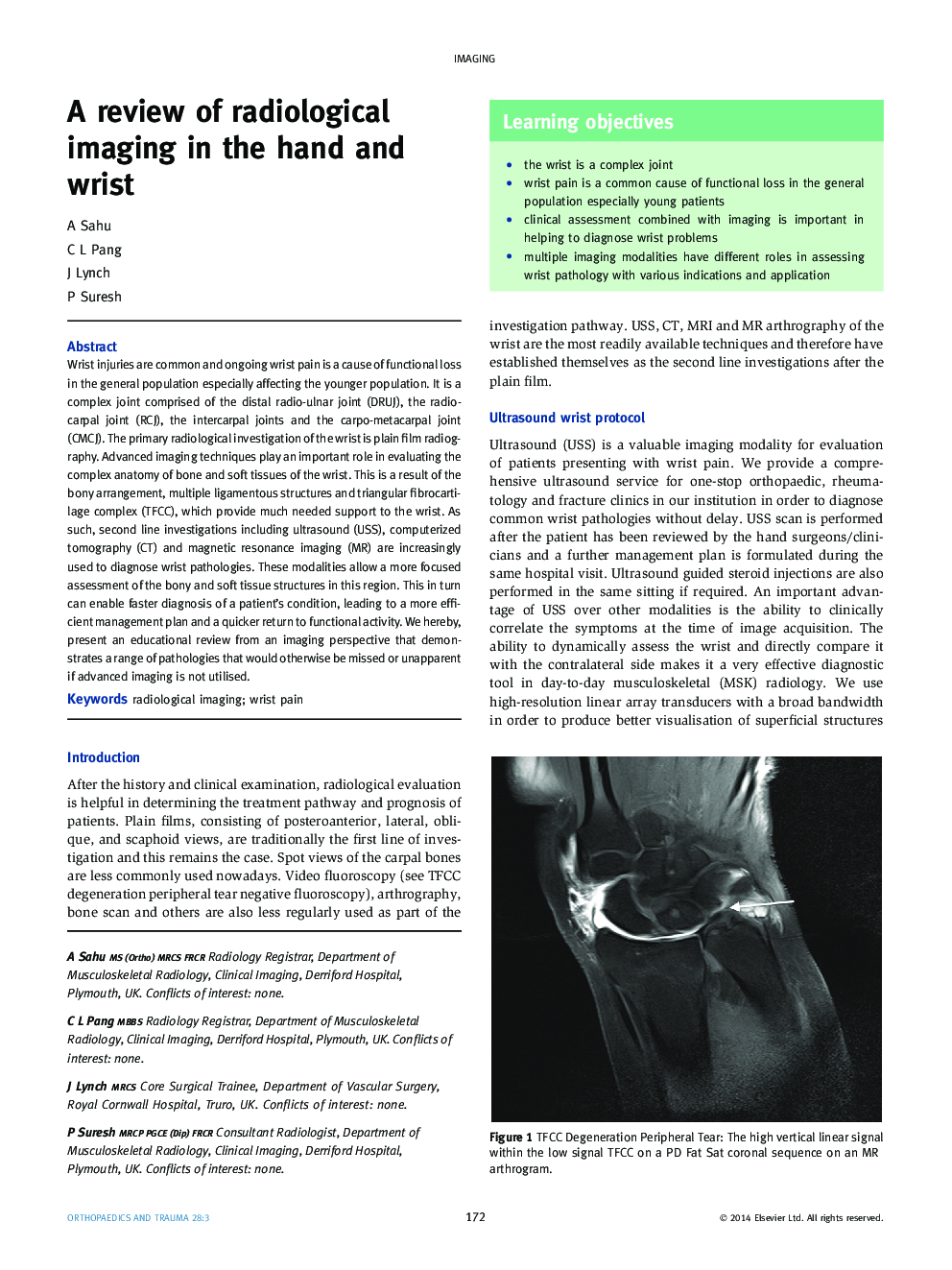
Wrist injuries are common and ongoing wrist pain is a cause of functional loss in the general population especially affecting the younger population. It is a complex joint comprised of the distal radio-ulnar joint (DRUJ), the radio-carpal joint (RCJ), the intercarpal joints and the carpo-metacarpal joint (CMCJ). The primary radiological investigation of the wrist is plain film radiography. Advanced imaging techniques play an important role in evaluating the complex anatomy of bone and soft tissues of the wrist. This is a result of the bony arrangement, multiple ligamentous structures and triangular fibrocartilage complex (TFCC), which provide much needed support to the wrist. As such, second line investigations including ultrasound (USS), computerized tomography (CT) and magnetic resonance imaging (MR) are increasingly used to diagnose wrist pathologies. These modalities allow a more focused assessment of the bony and soft tissue structures in this region. This in turn can enable faster diagnosis of a patient's condition, leading to a more efficient management plan and a quicker return to functional activity. We hereby, present an educational review from an imaging perspective that demonstrates a range of pathologies that would otherwise be missed or unapparent if advanced imaging is not utilised.
Journal: Orthopaedics and Trauma - Volume 28, Issue 3, June 2014, Pages 172–186
