| کد مقاله | کد نشریه | سال انتشار | مقاله انگلیسی | نسخه تمام متن |
|---|---|---|---|---|
| 4248326 | 1283731 | 2009 | 6 صفحه PDF | دانلود رایگان |
عنوان انگلیسی مقاله ISI
Giant recurrent glomus jugulotympanicum with intracranial, extracranial, and nasophayngeal extension: The imaging role in clinical management
دانلود مقاله + سفارش ترجمه
دانلود مقاله ISI انگلیسی
رایگان برای ایرانیان
کلمات کلیدی
موضوعات مرتبط
علوم پزشکی و سلامت
پزشکی و دندانپزشکی
رادیولوژی و تصویربرداری
پیش نمایش صفحه اول مقاله

چکیده انگلیسی
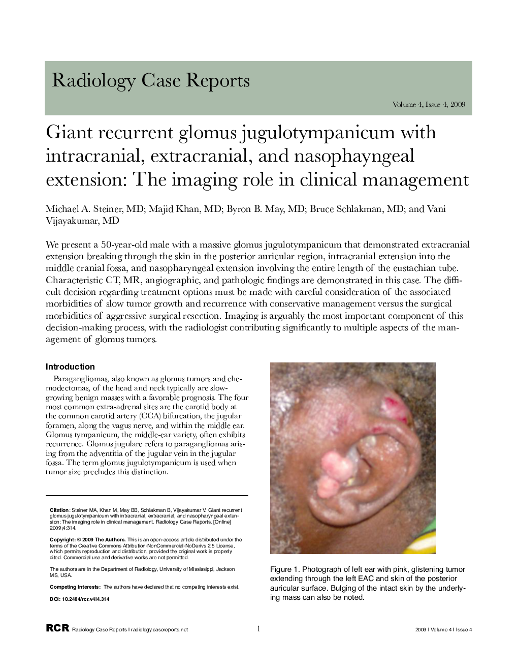
We present a 50-year-old male with a massive glomus jugulotympanicum that demonstrated extracranial extension breaking through the skin in the posterior auricular region, intracranial extension into the middle cranial fossa, and nasopharyngeal extension involving the entire length of the eustachian tube. Characteristic CT, MR, angiographic, and pathologic findings are demonstrated in this case. The difficult decision regarding treatment options must be made with careful consideration of the associated morbidities of slow tumor growth and recurrence with conservative management versus the surgical morbidities of aggressive surgical resection. Imaging is arguably the most important component of this decision-making process, with the radiologist contributing significantly to multiple aspects of the management of glomus tumors.
ناشر
Database: Elsevier - ScienceDirect (ساینس دایرکت)
Journal: Radiology Case Reports - Volume 4, Issue 4, 2009, 314
Journal: Radiology Case Reports - Volume 4, Issue 4, 2009, 314
نویسندگان
Michael A. MD, Majid MD, Byron B. MD, Bruce MD, Vani MD,