| کد مقاله | کد نشریه | سال انتشار | مقاله انگلیسی | نسخه تمام متن |
|---|---|---|---|---|
| 4259422 | 1284571 | 2010 | 7 صفحه PDF | دانلود رایگان |

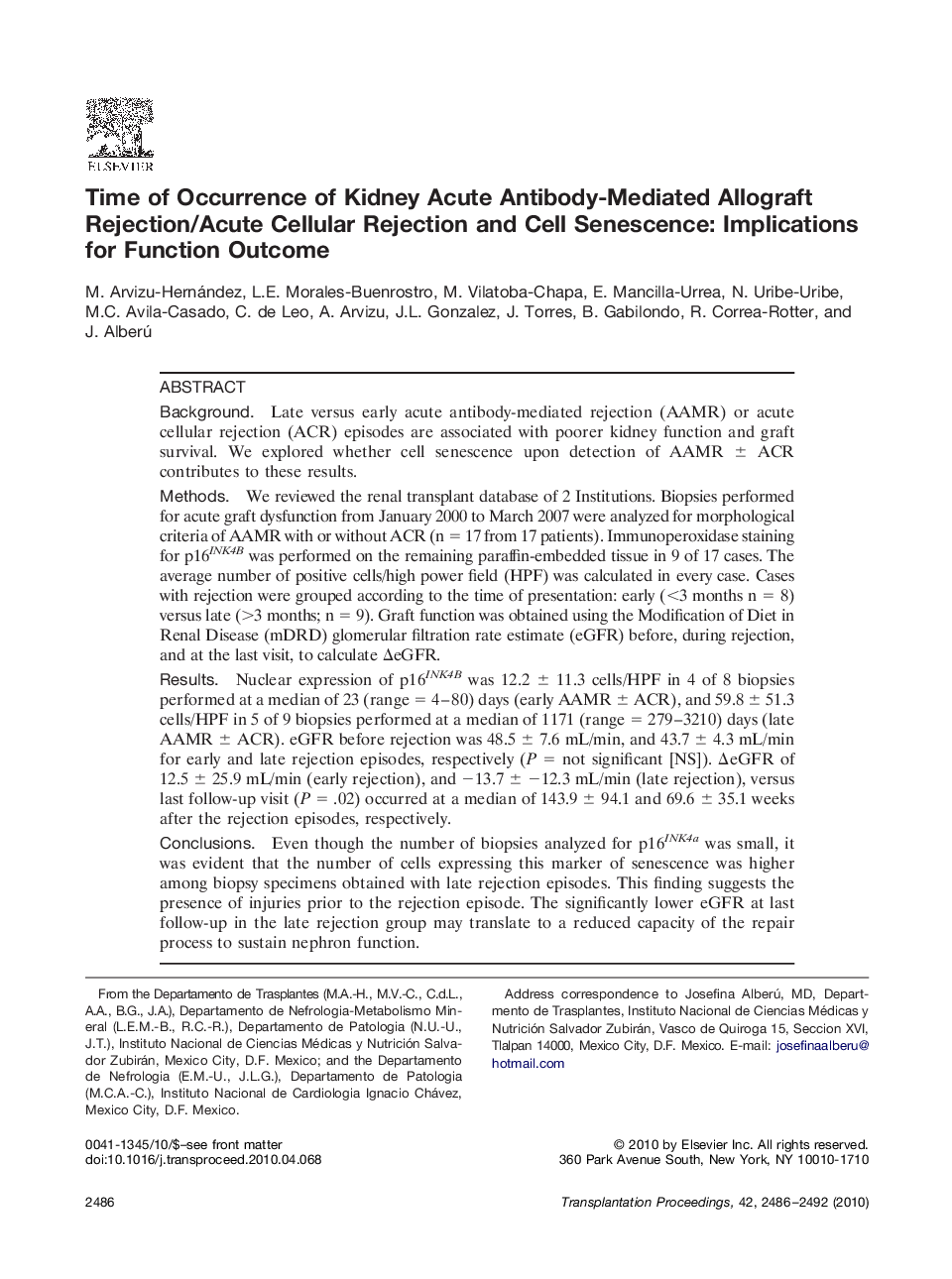
BackgroundLate versus early acute antibody-mediated rejection (AAMR) or acute cellular rejection (ACR) episodes are associated with poorer kidney function and graft survival. We explored whether cell senescence upon detection of AAMR ± ACR contributes to these results.MethodsWe reviewed the renal transplant database of 2 Institutions. Biopsies performed for acute graft dysfunction from January 2000 to March 2007 were analyzed for morphological criteria of AAMR with or without ACR (n = 17 from 17 patients). Immunoperoxidase staining for p16INK4B was performed on the remaining paraffin-embedded tissue in 9 of 17 cases. The average number of positive cells/high power field (HPF) was calculated in every case. Cases with rejection were grouped according to the time of presentation: early (<3 months n = 8) versus late (>3 months; n = 9). Graft function was obtained using the Modification of Diet in Renal Disease (mDRD) glomerular filtration rate estimate (eGFR) before, during rejection, and at the last visit, to calculate ΔeGFR.ResultsNuclear expression of p16INK4B was 12.2 ± 11.3 cells/HPF in 4 of 8 biopsies performed at a median of 23 (range = 4–80) days (early AAMR ± ACR), and 59.8 ± 51.3 cells/HPF in 5 of 9 biopsies performed at a median of 1171 (range = 279–3210) days (late AAMR ± ACR). eGFR before rejection was 48.5 ± 7.6 mL/min, and 43.7 ± 4.3 mL/min for early and late rejection episodes, respectively (P = not significant [NS]). ΔeGFR of 12.5 ± 25.9 mL/min (early rejection), and −13.7 ± −12.3 mL/min (late rejection), versus last follow-up visit (P = .02) occurred at a median of 143.9 ± 94.1 and 69.6 ± 35.1 weeks after the rejection episodes, respectively.ConclusionsEven though the number of biopsies analyzed for p16INK4a was small, it was evident that the number of cells expressing this marker of senescence was higher among biopsy specimens obtained with late rejection episodes. This finding suggests the presence of injuries prior to the rejection episode. The significantly lower eGFR at last follow-up in the late rejection group may translate to a reduced capacity of the repair process to sustain nephron function.
Journal: Transplantation Proceedings - Volume 42, Issue 7, September 2010, Pages 2486–2492