| کد مقاله | کد نشریه | سال انتشار | مقاله انگلیسی | نسخه تمام متن |
|---|---|---|---|---|
| 4341402 | 1295833 | 2007 | 19 صفحه PDF | دانلود رایگان |
عنوان انگلیسی مقاله ISI
A chronic histopathological and electrophysiological analysis of a rodent hypoxic-ischemic brain injury model and its use as a model of epilepsy
دانلود مقاله + سفارش ترجمه
دانلود مقاله ISI انگلیسی
رایگان برای ایرانیان
کلمات کلیدی
HEPESEGTAdl-2-amino-5-phosphonovaleric acidSNKIMLAP-5DNQXN-2-hydroxyethylpiperazine-N′-2-ethanesulfonic acid - N-2-hydroxyethylpiperazine-N'-2-ethanesulfonic acidEpilepsy - بیماری صرعRecurrent excitation - تحریک مجددanalysis of variance - تحلیل واریانسANOVA - تحلیل واریانس Analysis of varianceSprouting - جوانه زدنStudent-Newman-Keuls - دانش آموز-نیومن-کولpostnatal day - روز پس از زایمانPopulation spikes - شمارش جمعیتinner molecular layer - لایه داخلی مولکولیhypoxia-ischemia - هیپوکسی-ایسکمی
موضوعات مرتبط
علوم زیستی و بیوفناوری
علم عصب شناسی
علوم اعصاب (عمومی)
پیش نمایش صفحه اول مقاله

چکیده انگلیسی
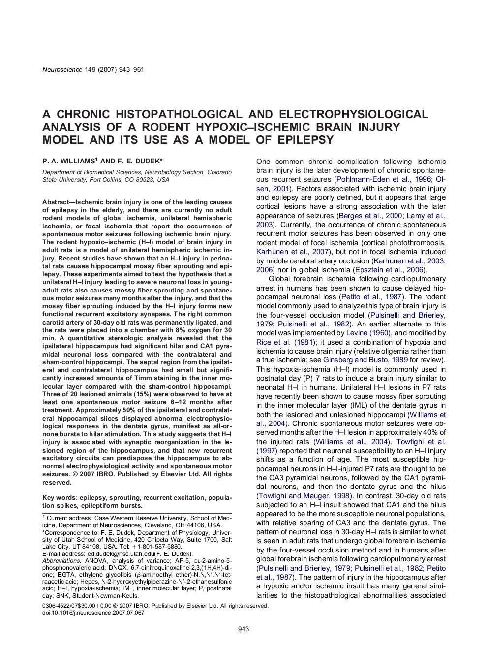
Ischemic brain injury is one of the leading causes of epilepsy in the elderly, and there are currently no adult rodent models of global ischemia, unilateral hemispheric ischemia, or focal ischemia that report the occurrence of spontaneous motor seizures following ischemic brain injury. The rodent hypoxic-ischemic (H-I) model of brain injury in adult rats is a model of unilateral hemispheric ischemic injury. Recent studies have shown that an H-I injury in perinatal rats causes hippocampal mossy fiber sprouting and epilepsy. These experiments aimed to test the hypothesis that a unilateral H-I injury leading to severe neuronal loss in young-adult rats also causes mossy fiber sprouting and spontaneous motor seizures many months after the injury, and that the mossy fiber sprouting induced by the H-I injury forms new functional recurrent excitatory synapses. The right common carotid artery of 30-day old rats was permanently ligated, and the rats were placed into a chamber with 8% oxygen for 30 min. A quantitative stereologic analysis revealed that the ipsilateral hippocampus had significant hilar and CA1 pyramidal neuronal loss compared with the contralateral and sham-control hippocampi. The septal region from the ipsilateral and contralateral hippocampus had small but significantly increased amounts of Timm staining in the inner molecular layer compared with the sham-control hippocampi. Three of 20 lesioned animals (15%) were observed to have at least one spontaneous motor seizure 6-12 months after treatment. Approximately 50% of the ipsilateral and contralateral hippocampal slices displayed abnormal electrophysiological responses in the dentate gyrus, manifest as all-or-none bursts to hilar stimulation. This study suggests that H-I injury is associated with synaptic reorganization in the lesioned region of the hippocampus, and that new recurrent excitatory circuits can predispose the hippocampus to abnormal electrophysiological activity and spontaneous motor seizures.
ناشر
Database: Elsevier - ScienceDirect (ساینس دایرکت)
Journal: Neuroscience - Volume 149, Issue 4, 23 November 2007, Pages 943-961
Journal: Neuroscience - Volume 149, Issue 4, 23 November 2007, Pages 943-961
نویسندگان
P.A. Williams, F.E. Dudek,