| کد مقاله | کد نشریه | سال انتشار | مقاله انگلیسی | نسخه تمام متن |
|---|---|---|---|---|
| 4341613 | 1295841 | 2008 | 10 صفحه PDF | دانلود رایگان |
عنوان انگلیسی مقاله ISI
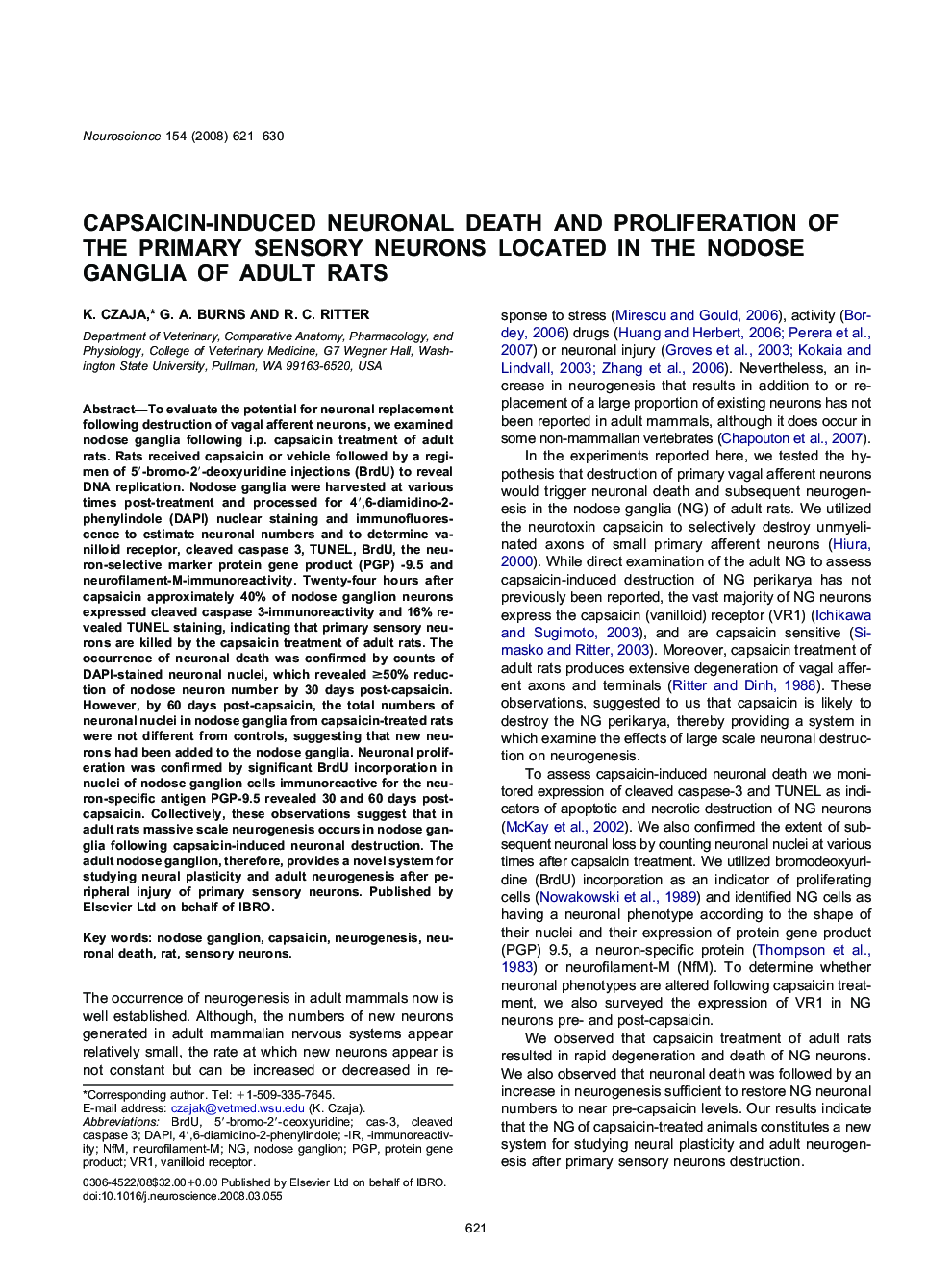
Capsaicin-induced neuronal death and proliferation of the primary sensory neurons located in the nodose ganglia of adult rats
دانلود مقاله + سفارش ترجمه
دانلود مقاله ISI انگلیسی
رایگان برای ایرانیان
کلمات کلیدی
-immunoreactivityVR1NFMDAPIPGP4′,6-diamidino-2-phenylindole - 4 '، 6-دیامیدینو-2-فنیلینول5′-bromo-2′-deoxyuridine - 5'-bromo-2'-deoxyuridineBrdU - بروموداکسی اوریدینcleaved caspase 3 - مخروطی شکسته 3Neuronal death - مرگ نورونcas-3 - مورد 3Rat - موش صحراییSensory neurons - نورونهای حسیNeurogenesis - نوروژنز-ir - وprotein gene product - پروتئین محصول ژنCapsaicin - کپسایسین یا کاپسیسینNodose ganglion - گنگلیون بینیVanilloid receptor - گیرنده وانیلیوئید
موضوعات مرتبط
علوم زیستی و بیوفناوری
علم عصب شناسی
علوم اعصاب (عمومی)
پیش نمایش صفحه اول مقاله

چکیده انگلیسی
To evaluate the potential for neuronal replacement following destruction of vagal afferent neurons, we examined nodose ganglia following i.p. capsaicin treatment of adult rats. Rats received capsaicin or vehicle followed by a regimen of 5â²-bromo-2â²-deoxyuridine injections (BrdU) to reveal DNA replication. Nodose ganglia were harvested at various times post-treatment and processed for 4â²,6-diamidino-2-phenylindole (DAPI) nuclear staining and immunofluorescence to estimate neuronal numbers and to determine vanilloid receptor, cleaved caspase 3, TUNEL, BrdU, the neuron-selective marker protein gene product (PGP) -9.5 and neurofilament-M-immunoreactivity. Twenty-four hours after capsaicin approximately 40% of nodose ganglion neurons expressed cleaved caspase 3-immunoreactivity and 16% revealed TUNEL staining, indicating that primary sensory neurons are killed by the capsaicin treatment of adult rats. The occurrence of neuronal death was confirmed by counts of DAPI-stained neuronal nuclei, which revealed â¥50% reduction of nodose neuron number by 30 days post-capsaicin. However, by 60 days post-capsaicin, the total numbers of neuronal nuclei in nodose ganglia from capsaicin-treated rats were not different from controls, suggesting that new neurons had been added to the nodose ganglia. Neuronal proliferation was confirmed by significant BrdU incorporation in nuclei of nodose ganglion cells immunoreactive for the neuron-specific antigen PGP-9.5 revealed 30 and 60 days post-capsaicin. Collectively, these observations suggest that in adult rats massive scale neurogenesis occurs in nodose ganglia following capsaicin-induced neuronal destruction. The adult nodose ganglion, therefore, provides a novel system for studying neural plasticity and adult neurogenesis after peripheral injury of primary sensory neurons.
ناشر
Database: Elsevier - ScienceDirect (ساینس دایرکت)
Journal: Neuroscience - Volume 154, Issue 2, 23 June 2008, Pages 621-630
Journal: Neuroscience - Volume 154, Issue 2, 23 June 2008, Pages 621-630
نویسندگان
K. Czaja, G.A. Burns, R.C. Ritter,