| کد مقاله | کد نشریه | سال انتشار | مقاله انگلیسی | نسخه تمام متن |
|---|---|---|---|---|
| 4356245 | 1615674 | 2007 | 10 صفحه PDF | دانلود رایگان |
عنوان انگلیسی مقاله ISI
Distribution of focal lesions in the chinchilla organ of Corti following exposure to a 4-kHz or a 0.5-kHz octave band of noise
دانلود مقاله + سفارش ترجمه
دانلود مقاله ISI انگلیسی
رایگان برای ایرانیان
کلمات کلیدی
RNSNIHLROS - ROSnoise-induced hearing loss - افت شنوایی ناشی از سر و صداorgan of Corti - اندام کورتیNoise - سر و صداFocal lesions - ضایعات کانونیbasilar membrane - غشاء basilarCharacteristic Frequency - فرکانس مشخصهChinchilla - چینچیلاreactive nitrogen species - گونه های واکنش پذیر نیتروژنReactive oxygen species - گونههای فعال اکسیژن
موضوعات مرتبط
علوم زیستی و بیوفناوری
علم عصب شناسی
سیستم های حسی
پیش نمایش صفحه اول مقاله

چکیده انگلیسی
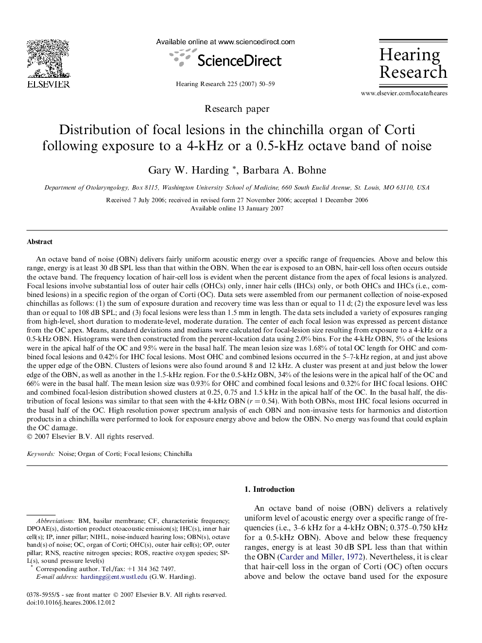
An octave band of noise (OBN) delivers fairly uniform acoustic energy over a specific range of frequencies. Above and below this range, energy is at least 30 dB SPL less than that within the OBN. When the ear is exposed to an OBN, hair-cell loss often occurs outside the octave band. The frequency location of hair-cell loss is evident when the percent distance from the apex of focal lesions is analyzed. Focal lesions involve substantial loss of outer hair cells (OHCs) only, inner hair cells (IHCs) only, or both OHCs and IHCs (i.e., combined lesions) in a specific region of the organ of Corti (OC). Data sets were assembled from our permanent collection of noise-exposed chinchillas as follows: (1) the sum of exposure duration and recovery time was less than or equal to 11 d; (2) the exposure level was less than or equal to 108 dB SPL; and (3) focal lesions were less than 1.5 mm in length. The data sets included a variety of exposures ranging from high-level, short duration to moderate-level, moderate duration. The center of each focal lesion was expressed as percent distance from the OC apex. Means, standard deviations and medians were calculated for focal-lesion size resulting from exposure to a 4-kHz or a 0.5-kHz OBN. Histograms were then constructed from the percent-location data using 2.0% bins. For the 4-kHz OBN, 5% of the lesions were in the apical half of the OC and 95% were in the basal half. The mean lesion size was 1.68% of total OC length for OHC and combined focal lesions and 0.42% for IHC focal lesions. Most OHC and combined lesions occurred in the 5-7-kHz region, at and just above the upper edge of the OBN. Clusters of lesions were also found around 8 and 12 kHz. A cluster was present at and just below the lower edge of the OBN, as well as another in the 1.5-kHz region. For the 0.5-kHz OBN, 34% of the lesions were in the apical half of the OC and 66% were in the basal half. The mean lesion size was 0.93% for OHC and combined focal lesions and 0.32% for IHC focal lesions. OHC and combined focal-lesion distribution showed clusters at 0.25, 0.75 and 1.5 kHz in the apical half of the OC. In the basal half, the distribution of focal lesions was similar to that seen with the 4-kHz OBN (r = 0.54). With both OBNs, most IHC focal lesions occurred in the basal half of the OC. High resolution power spectrum analysis of each OBN and non-invasive tests for harmonics and distortion products in a chinchilla were performed to look for exposure energy above and below the OBN. No energy was found that could explain the OC damage.
ناشر
Database: Elsevier - ScienceDirect (ساینس دایرکت)
Journal: Hearing Research - Volume 225, Issues 1â2, March 2007, Pages 50-59
Journal: Hearing Research - Volume 225, Issues 1â2, March 2007, Pages 50-59
نویسندگان
Gary W. Harding, Barbara A. Bohne,