| کد مقاله | کد نشریه | سال انتشار | مقاله انگلیسی | نسخه تمام متن |
|---|---|---|---|---|
| 5737623 | 1614718 | 2017 | 23 صفحه PDF | دانلود رایگان |

- Non-invasive imaging is an important tool for preclinical epilepsy research.
- Neuroimaging represents a translatable technique to identify early biomarkers.
- Longitudinal neuroimaging can improve our knowledge of the process of epileptogenesis.
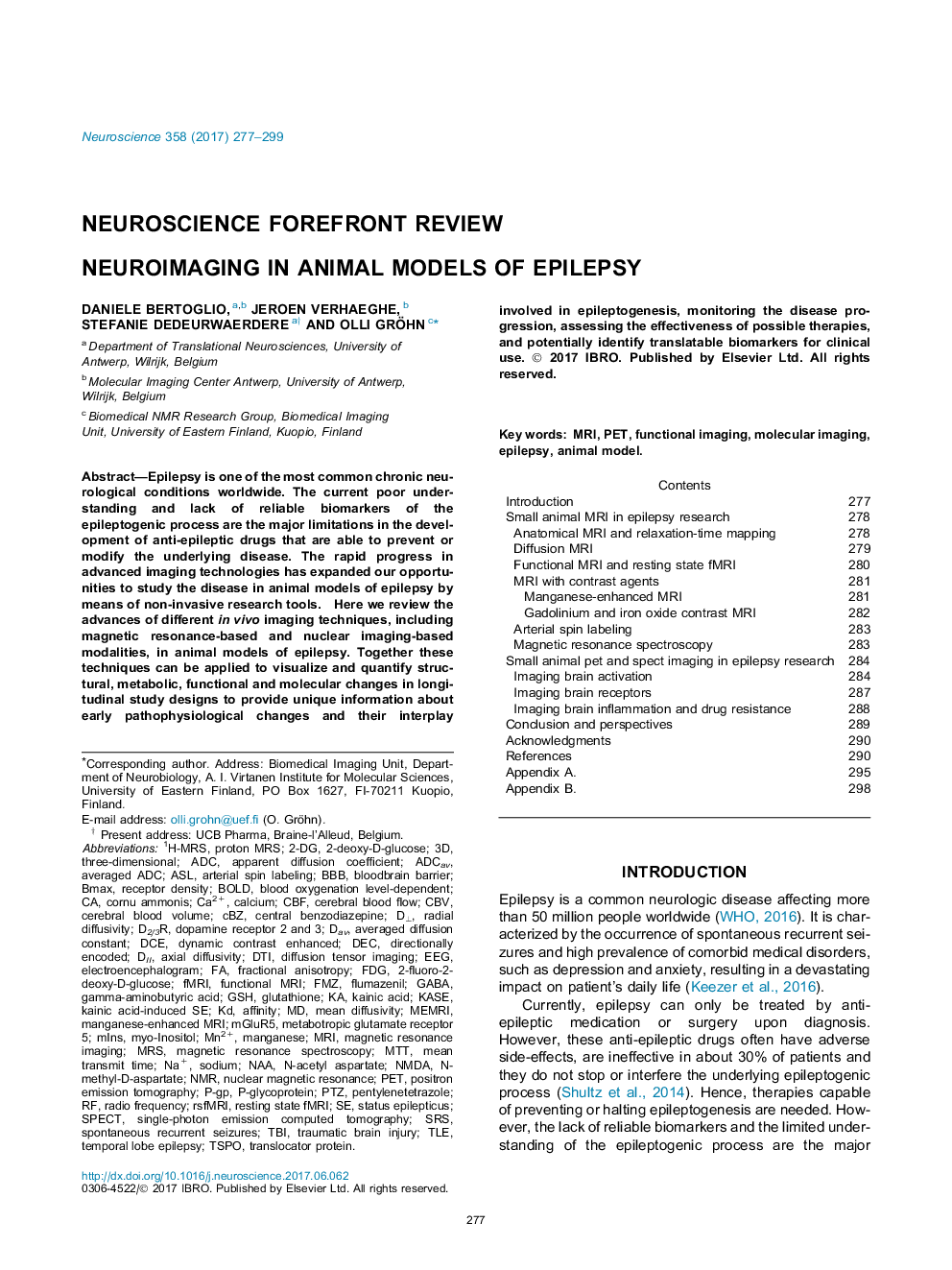
Epilepsy is one of the most common chronic neurological conditions worldwide. The current poor understanding and lack of reliable biomarkers of the epileptogenic process are the major limitations in the development of anti-epileptic drugs that are able to prevent or modify the underlying disease. The rapid progress in advanced imaging technologies has expanded our opportunities to study the disease in animal models of epilepsy by means of non-invasive research tools.Here we review the advances of different in vivo imaging techniques, including magnetic resonance-based and nuclear imaging-based modalities, in animal models of epilepsy. Together these techniques can be applied to visualize and quantify structural, metabolic, functional and molecular changes in longitudinal study designs to provide unique information about early pathophysiological changes and their interplay involved in epileptogenesis, monitoring the disease progression, assessing the effectiveness of possible therapies, and potentially identify translatable biomarkers for clinical use.
Journal: Neuroscience - Volume 358, 1 September 2017, Pages 277-299