| کد مقاله | کد نشریه | سال انتشار | مقاله انگلیسی | نسخه تمام متن |
|---|---|---|---|---|
| 6037048 | 1188783 | 2010 | 10 صفحه PDF | دانلود رایگان |
عنوان انگلیسی مقاله ISI
Feature-based morphometry: Discovering group-related anatomical patterns
دانلود مقاله + سفارش ترجمه
دانلود مقاله ISI انگلیسی
رایگان برای ایرانیان
کلمات کلیدی
موضوعات مرتبط
علوم زیستی و بیوفناوری
علم عصب شناسی
علوم اعصاب شناختی
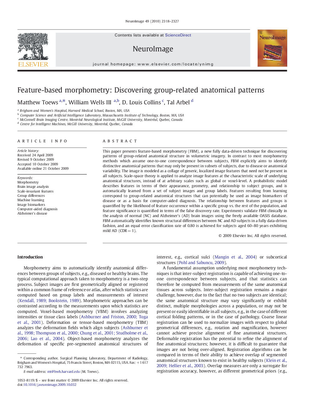
پیش نمایش صفحه اول مقاله

چکیده انگلیسی
This paper presents feature-based morphometry (FBM), a new fully data-driven technique for discovering patterns of group-related anatomical structure in volumetric imagery. In contrast to most morphometry methods which assume one-to-one correspondence between subjects, FBM explicitly aims to identify distinctive anatomical patterns that may only be present in subsets of subjects, due to disease or anatomical variability. The image is modeled as a collage of generic, localized image features that need not be present in all subjects. Scale-space theory is applied to analyze image features at the characteristic scale of underlying anatomical structures, instead of at arbitrary scales such as global or voxel-level. A probabilistic model describes features in terms of their appearance, geometry, and relationship to subject groups, and is automatically learned from a set of subject images and group labels. Features resulting from learning correspond to group-related anatomical structures that can potentially be used as image biomarkers of disease or as a basis for computer-aided diagnosis. The relationship between features and groups is quantified by the likelihood of feature occurrence within a specific group vs. the rest of the population, and feature significance is quantified in terms of the false discovery rate. Experiments validate FBM clinically in the analysis of normal (NC) and Alzheimer's (AD) brain images using the freely available OASIS database. FBM automatically identifies known structural differences between NC and AD subjects in a fully data-driven fashion, and an equal error classification rate of 0.80 is achieved for subjects aged 60-80 years exhibiting mild AD (CDRÂ =Â 1).
ناشر
Database: Elsevier - ScienceDirect (ساینس دایرکت)
Journal: NeuroImage - Volume 49, Issue 3, 1 February 2010, Pages 2318-2327
Journal: NeuroImage - Volume 49, Issue 3, 1 February 2010, Pages 2318-2327
نویسندگان
Matthew Toews, William III, D. Louis Collins, Tal Arbel,