| کد مقاله | کد نشریه | سال انتشار | مقاله انگلیسی | نسخه تمام متن |
|---|---|---|---|---|
| 6267579 | 1614599 | 2016 | 8 صفحه PDF | دانلود رایگان |
- Semi-automated methodology utilizes fluorescence microscopy to calculate vascular density within brain and brain tumors.
- Novel methodology produces data comparable to previously hand counting pathology methods.
- Vascular density is decreased in experimental brain metastases compared to a preclinical glioma model.
BackgroundMeasurement of vascular density has significant value in characterizing healthy and diseased tissue, particularly in brain where vascular density varies among regions. Further, an understanding of brain vessel size helps distinguish between capillaries and larger vessels like arterioles and venules. Unfortunately, few cutting edge methodologies are available to laboratories to rapidly quantify vessel density.New methodWe developed a rapid microscopic method, which quantifies the numbers and diameters of blood vessels in brain. Utilizing this method we characterized vascular density of five brain regions in both mice and rats, in two tumor models, using three tracers.ResultsWe observed the number of sections/mm2 in various brain regions: genu of corpus callosum 161 ± 7, hippocampus 266 ± 18, superior colliculus 300 ± 24, frontal cortex 391 ± 55, and inferior colliculus 692 ± 18 (n = 5 animals). Regional brain data were not significantly different between species (p > 0.05) or when using different tracers (70 kDa and 2000 kDa Texas Red; p > 0.05). Vascular density decreased (62-79%) in preclinical brain metastases but increased (62%) a rat glioma model.Comparison with existing methodsOur values were similar (p > 0.05) to published literature. We applied this method to brain-tumors and observed brain metastases of breast cancer to have a â¼2.5-fold reduction (p > 0.05) in vessels/mm2 compared to normal cortical regions. In contrast, vascular density in a glioma model was significantly higher (sections/mm2 736 ± 84; p < 0.05).ConclusionsIn summary, we present a vascular density counting method that is rapid, sensitive, and uses fluorescence microscopy without antibodies.
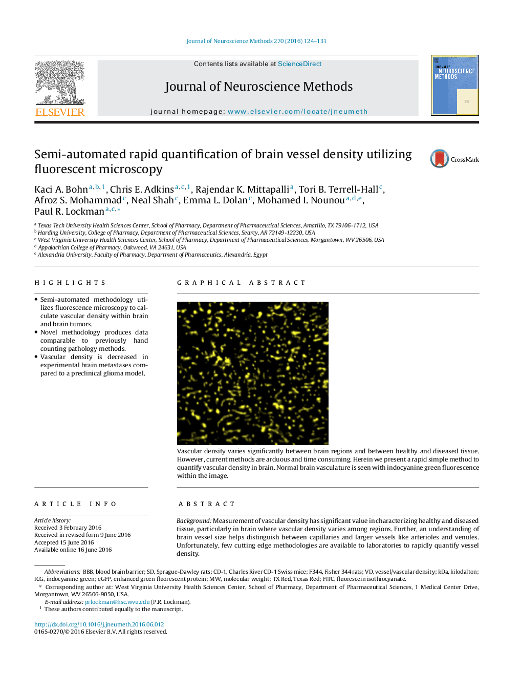
151Vascular density varies significantly between brain regions and between healthy and diseased tissue. However, current methods are arduous and time consuming. Herein we present a rapid simple method to quantify vascular density in brain. Normal brain vasculature is seen with indocyanine green fluorescence within the image.
Journal: Journal of Neuroscience Methods - Volume 270, 1 September 2016, Pages 124-131
