| کد مقاله | کد نشریه | سال انتشار | مقاله انگلیسی | نسخه تمام متن |
|---|---|---|---|---|
| 6272975 | 1614792 | 2015 | 14 صفحه PDF | دانلود رایگان |
عنوان انگلیسی مقاله ISI
Increased levels and activity of cathepsins B and D in kainate-induced toxicity
دانلود مقاله + سفارش ترجمه
دانلود مقاله ISI انگلیسی
رایگان برای ایرانیان
کلمات کلیدی
HRPBCAECLTLEinsulin-like growth factor-II receptorAIF - آیفونenhanced chemiluminescence - بهبود شیمیایی لومنbicinchoninic acid - بیسینکنینیک اسیدEpilepsy - بیماری صرعSeizures - تشنجNeurodegeneration - تولید نوروژنیکtemporal lobe epilepsy - صرع لوب تمپورالapoptosis-inducing factor - عامل القاء آپوپتوزHippocampus - هیپوکامپ Horseradish peroxidase - پراکسیداز هوررادیشCathepsins - کاتهپسین ها
موضوعات مرتبط
علوم زیستی و بیوفناوری
علم عصب شناسی
علوم اعصاب (عمومی)
پیش نمایش صفحه اول مقاله

چکیده انگلیسی
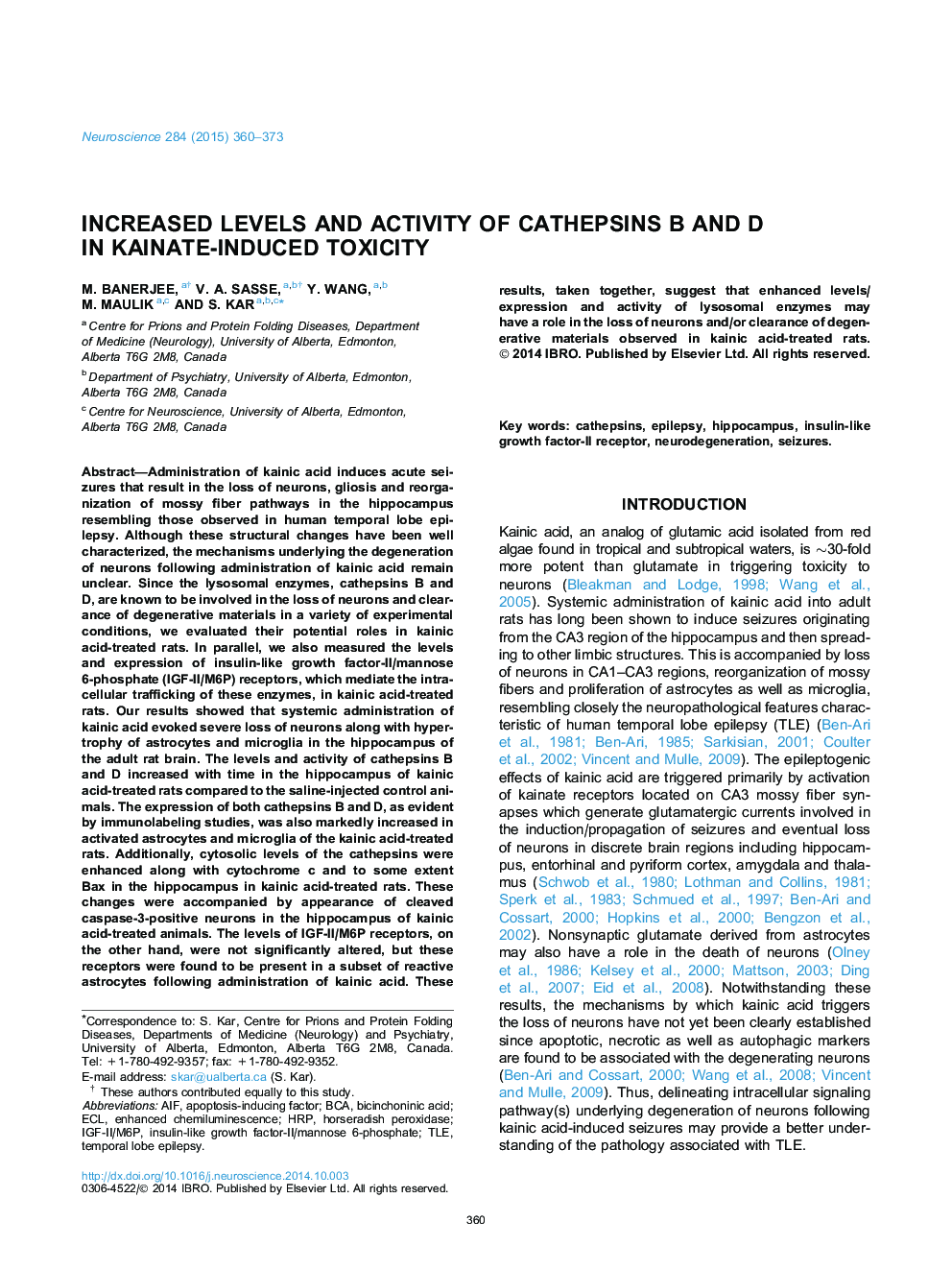
Administration of kainic acid induces acute seizures that result in the loss of neurons, gliosis and reorganization of mossy fiber pathways in the hippocampus resembling those observed in human temporal lobe epilepsy. Although these structural changes have been well characterized, the mechanisms underlying the degeneration of neurons following administration of kainic acid remain unclear. Since the lysosomal enzymes, cathepsins B and D, are known to be involved in the loss of neurons and clearance of degenerative materials in a variety of experimental conditions, we evaluated their potential roles in kainic acid-treated rats. In parallel, we also measured the levels and expression of insulin-like growth factor-II/mannose 6-phosphate (IGF-II/M6P) receptors, which mediate the intracellular trafficking of these enzymes, in kainic acid-treated rats. Our results showed that systemic administration of kainic acid evoked severe loss of neurons along with hypertrophy of astrocytes and microglia in the hippocampus of the adult rat brain. The levels and activity of cathepsins B and D increased with time in the hippocampus of kainic acid-treated rats compared to the saline-injected control animals. The expression of both cathepsins B and D, as evident by immunolabeling studies, was also markedly increased in activated astrocytes and microglia of the kainic acid-treated rats. Additionally, cytosolic levels of the cathepsins were enhanced along with cytochrome c and to some extent Bax in the hippocampus in kainic acid-treated rats. These changes were accompanied by appearance of cleaved caspase-3-positive neurons in the hippocampus of kainic acid-treated animals. The levels of IGF-II/M6P receptors, on the other hand, were not significantly altered, but these receptors were found to be present in a subset of reactive astrocytes following administration of kainic acid. These results, taken together, suggest that enhanced levels/expression and activity of lysosomal enzymes may have a role in the loss of neurons and/or clearance of degenerative materials observed in kainic acid-treated rats.
ناشر
Database: Elsevier - ScienceDirect (ساینس دایرکت)
Journal: Neuroscience - Volume 284, 22 January 2015, Pages 360-373
Journal: Neuroscience - Volume 284, 22 January 2015, Pages 360-373
نویسندگان
M. Banerjee, V.A. Sasse, Y. Wang, M. Maulik, S. Kar,