| کد مقاله | کد نشریه | سال انتشار | مقاله انگلیسی | نسخه تمام متن |
|---|---|---|---|---|
| 7704941 | 1496903 | 2016 | 10 صفحه PDF | دانلود رایگان |
عنوان انگلیسی مقاله ISI
Dielectric relaxations on erythrocyte membrane as revealed by spectrin denaturation
دانلود مقاله + سفارش ترجمه
دانلود مقاله ISI انگلیسی
رایگان برای ایرانیان
کلمات کلیدی
موضوعات مرتبط
مهندسی و علوم پایه
شیمی
الکتروشیمی
پیش نمایش صفحه اول مقاله

چکیده انگلیسی
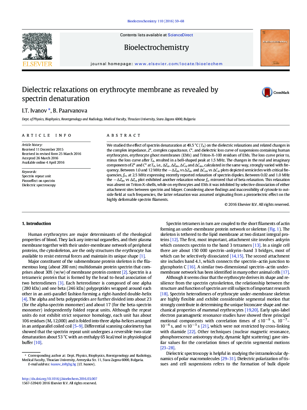
We studied the effect of spectrin denaturation at 49.5 °C (TA) on the dielectric relaxations and related changes in the complex impedance, Z*, complex capacitance, C*, and dielectric loss curve of suspensions containing human erythrocytes, erythrocyte ghost membranes (EMs) and Triton-X-100 residues of EMs. The loss curve prior to, minus the loss curve after TA, resulted in a bell-shaped peak at 1.5 MHz. The changes in the real and imaginary components of Z* and C* at TA, i.e., ÎZre, ÎZim, ÎCre and ÎCim, calculated in the same way, strongly varied with frequency. Between 1.0 and 12 MHz the â ÎZim vs ÎZre, and ÎCim vs ÎCre plots depicted semicircles with critical frequencies, fcr, at 2.5 MHz expressing recently reported relaxation of spectrin dipoles. Between 0.02 and 1.0 MHz the â ÎZim vs ÎZre plot exhibited another relaxation whose fcr mirrored that of beta relaxation. This relaxation was absent on Triton-X-shells, while on erythrocytes and EMs it was inhibited by selective dissociation of either attachment sites between spectrin and bilayer. Considering above findings and inaccessibility of cytosole to outside field at such frequencies, the latter relaxation was assumed originating from a piezoelectric effect on the highly deformable spectrin filaments.
ناشر
Database: Elsevier - ScienceDirect (ساینس دایرکت)
Journal: Bioelectrochemistry - Volume 110, August 2016, Pages 59-68
Journal: Bioelectrochemistry - Volume 110, August 2016, Pages 59-68
نویسندگان
I.T. Ivanov, B. Paarvanova,