| کد مقاله | کد نشریه | سال انتشار | مقاله انگلیسی | نسخه تمام متن |
|---|---|---|---|---|
| 7925075 | 1512501 | 2018 | 9 صفحه PDF | دانلود رایگان |
عنوان انگلیسی مقاله ISI
Review on methods of solving the refractive index-thickness coupling problem in digital holographic microscopy of biological cells
ترجمه فارسی عنوان
بررسی روش حل مساله ضریب شکست ضخامت در میکروسکوپ هولوگرافی دیجیتال سلول های بیولوژیکی
دانلود مقاله + سفارش ترجمه
دانلود مقاله ISI انگلیسی
رایگان برای ایرانیان
کلمات کلیدی
میکروسکوپ هولوگرافی دیجیتال، میکروسکوپ فازی کمی، میکروسکوپ فازی اینترفرومتریک، جدا کردن، میکروسکوپ فیزیکی توموگرافی، ضریب شکست،
ترجمه چکیده
میکروسکوپ هولوگرافی دیجیتال یک روش تصویربرداری پر رونق است که علاقمندی قابل توجه تحقیق به تصویربرداری سلول های بیولوژیکی کمی را به دلیل توانایی آن برای ایجاد کنتراست بدون برچسب بدون علامت ایجاد می کند، بلکه اطلاعات فیزیکی بالایی را در رابطه با تراکم و ابعاد نمونه با مقیاس نانومتری ارائه می دهد حساسیت محوری این تکنیک الگوی تداخل بین پرتو نمونه و یک پرتو مرجع را ثبت می کند و با پردازش دیجیتال آن، می تواند تاخیر مسیر نوری بین این پرتوها را بازسازی کند. در هر نقطه فضایی، نقشه تاخیر مسیر نوری متناسب با محصول ضخامت فیزیکی نمونه و شاخص شکست انحلال نمونه است. از آنجایی که شاخص انکسار سلول محتویات آن را بدون نیاز به برچسب گذاری نشان می دهد، بسیار مفید است که ضخامت فیزیکی سلول را از مشخصات شاخص انکسار آن جدا کنید. این مقاله به بررسی روش های مختلف استخراج شاخص انکسار از اندازه گیری های میکروسکوپ های هولوگرافی دیجیتال سلول ها می پردازد. به محض اینکه شاخص انکسار سلول در دسترس باشد، می توان آن را برای هر دو آزمایش های بیولوژیکی یا تشخیص پزشکی مورد استفاده قرار داد، همانطور که در این دستنوشته بررسی شده است.
موضوعات مرتبط
مهندسی و علوم پایه
مهندسی مواد
مواد الکترونیکی، نوری و مغناطیسی
چکیده انگلیسی
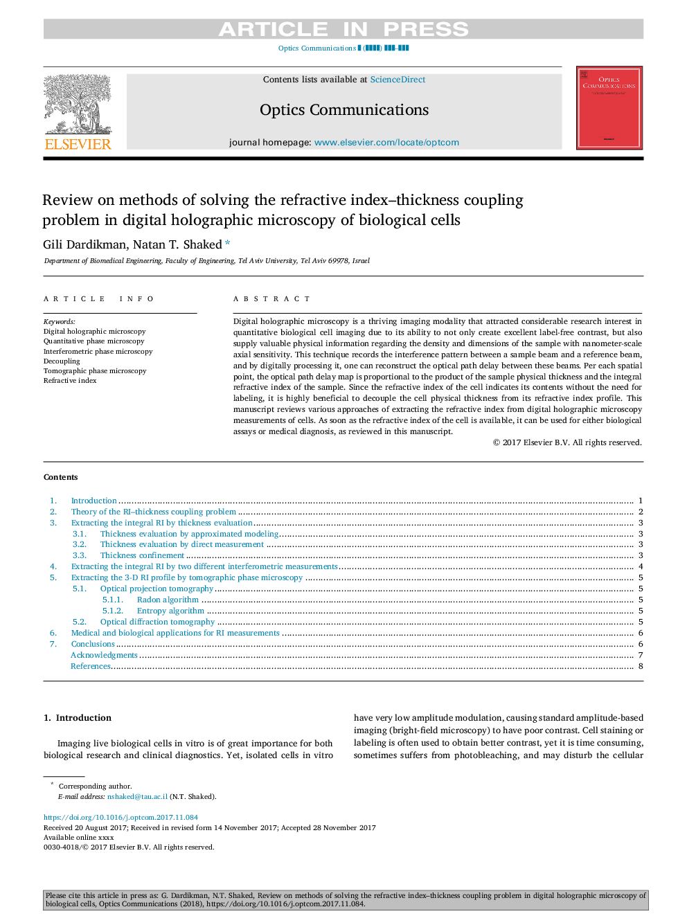
Digital holographic microscopy is a thriving imaging modality that attracted considerable research interest in quantitative biological cell imaging due to its ability to not only create excellent label-free contrast, but also supply valuable physical information regarding the density and dimensions of the sample with nanometer-scale axial sensitivity. This technique records the interference pattern between a sample beam and a reference beam, and by digitally processing it, one can reconstruct the optical path delay between these beams. Per each spatial point, the optical path delay map is proportional to the product of the sample physical thickness and the integral refractive index of the sample. Since the refractive index of the cell indicates its contents without the need for labeling, it is highly beneficial to decouple the cell physical thickness from its refractive index profile. This manuscript reviews various approaches of extracting the refractive index from digital holographic microscopy measurements of cells. As soon as the refractive index of the cell is available, it can be used for either biological assays or medical diagnosis, as reviewed in this manuscript.
ناشر
Database: Elsevier - ScienceDirect (ساینس دایرکت)
Journal: Optics Communications - Volume 422, 1 September 2018, Pages 8-16
Journal: Optics Communications - Volume 422, 1 September 2018, Pages 8-16
نویسندگان
Gili Dardikman, Natan T. Shaked,
