| کد مقاله | کد نشریه | سال انتشار | مقاله انگلیسی | نسخه تمام متن |
|---|---|---|---|---|
| 8697935 | 1584097 | 2017 | 8 صفحه PDF | دانلود رایگان |
عنوان انگلیسی مقاله ISI
Characterizing the skull base in craniofacial microsomia using principal component analysis
دانلود مقاله + سفارش ترجمه
دانلود مقاله ISI انگلیسی
رایگان برای ایرانیان
کلمات کلیدی
موضوعات مرتبط
علوم پزشکی و سلامت
پزشکی و دندانپزشکی
دندانپزشکی، جراحی دهان و پزشکی
پیش نمایش صفحه اول مقاله

چکیده انگلیسی
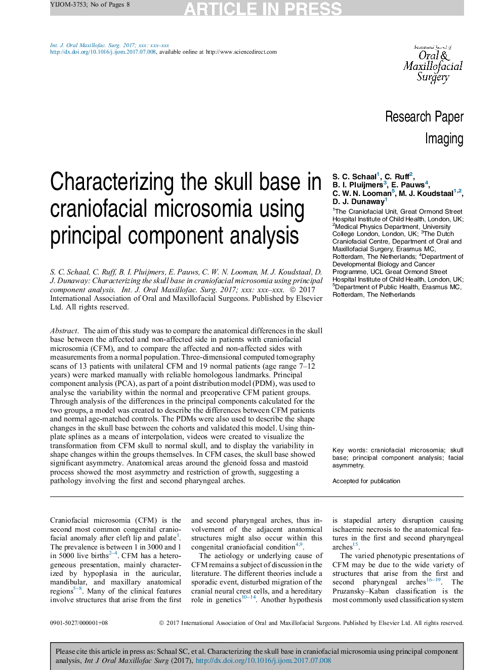
The aim of this study was to compare the anatomical differences in the skull base between the affected and non-affected side in patients with craniofacial microsomia (CFM), and to compare the affected and non-affected sides with measurements from a normal population. Three-dimensional computed tomography scans of 13 patients with unilateral CFM and 19 normal patients (age range 7-12 years) were marked manually with reliable homologous landmarks. Principal component analysis (PCA), as part of a point distribution model (PDM), was used to analyse the variability within the normal and preoperative CFM patient groups. Through analysis of the differences in the principal components calculated for the two groups, a model was created to describe the differences between CFM patients and normal age-matched controls. The PDMs were also used to describe the shape changes in the skull base between the cohorts and validated this model. Using thin-plate splines as a means of interpolation, videos were created to visualize the transformation from CFM skull to normal skull, and to display the variability in shape changes within the groups themselves. In CFM cases, the skull base showed significant asymmetry. Anatomical areas around the glenoid fossa and mastoid process showed the most asymmetry and restriction of growth, suggesting a pathology involving the first and second pharyngeal arches.
ناشر
Database: Elsevier - ScienceDirect (ساینس دایرکت)
Journal: International Journal of Oral and Maxillofacial Surgery - Volume 46, Issue 12, December 2017, Pages 1656-1663
Journal: International Journal of Oral and Maxillofacial Surgery - Volume 46, Issue 12, December 2017, Pages 1656-1663
نویسندگان
S.C. Schaal, C. Ruff, B.I. Pluijmers, E. Pauws, C.W.N. Looman, M.J. Koudstaal, D.J. Dunaway,