| کد مقاله | کد نشریه | سال انتشار | مقاله انگلیسی | نسخه تمام متن |
|---|---|---|---|---|
| 8719020 | 1588888 | 2017 | 7 صفحه PDF | دانلود رایگان |
عنوان انگلیسی مقاله ISI
When does intraoperative 3D-imaging play a role in transpedicular C2 screw placement?
دانلود مقاله + سفارش ترجمه
دانلود مقاله ISI انگلیسی
رایگان برای ایرانیان
کلمات کلیدی
موضوعات مرتبط
علوم پزشکی و سلامت
پزشکی و دندانپزشکی
طب اورژانس
پیش نمایش صفحه اول مقاله

چکیده انگلیسی
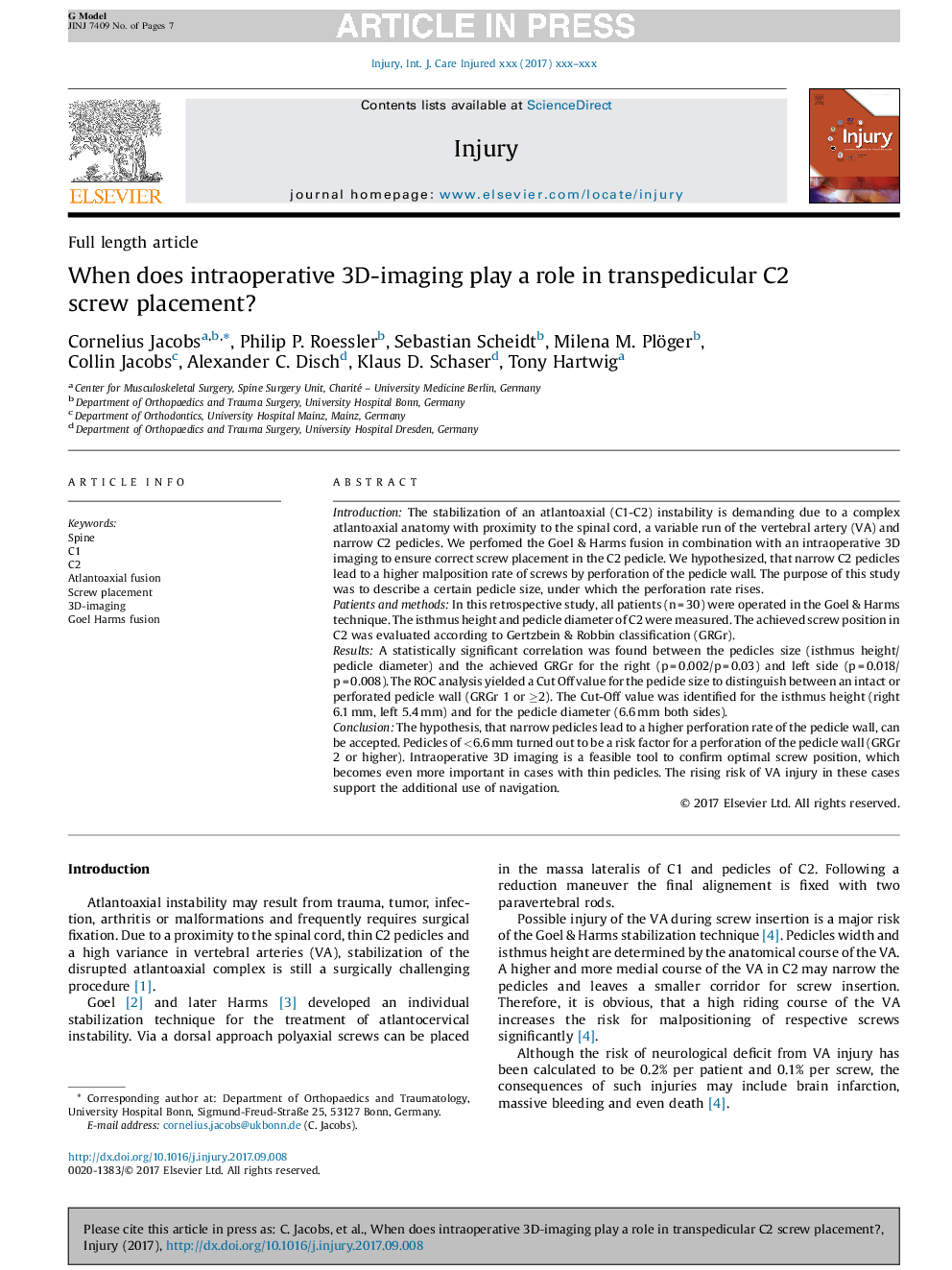
The hypothesis, that narrow pedicles lead to a higher perforation rate of the pedicle wall, can be accepted. Pedicles of <6.6Â mm turned out to be a risk factor for a perforation of the pedicle wall (GRGr 2 or higher). Intraoperative 3D imaging is a feasible tool to confirm optimal screw position, which becomes even more important in cases with thin pedicles. The rising risk of VA injury in these cases support the additional use of navigation.
ناشر
Database: Elsevier - ScienceDirect (ساینس دایرکت)
Journal: Injury - Volume 48, Issue 11, November 2017, Pages 2522-2528
Journal: Injury - Volume 48, Issue 11, November 2017, Pages 2522-2528
نویسندگان
Cornelius Jacobs, Philip P. Roessler, Sebastian Scheidt, Milena M. Plöger, Collin Jacobs, Alexander C. Disch, Klaus D. Schaser, Tony Hartwig,