| کد مقاله | کد نشریه | سال انتشار | مقاله انگلیسی | نسخه تمام متن |
|---|---|---|---|---|
| 8966714 | 1101258 | 2005 | 4 صفحه PDF | دانلود رایگان |
عنوان انگلیسی مقاله ISI
How to Radiograph the Erupted (Clinical) Crown of Equine Cheek Teeth
دانلود مقاله + سفارش ترجمه
دانلود مقاله ISI انگلیسی
رایگان برای ایرانیان
کلمات کلیدی
موضوعات مرتبط
علوم پزشکی و سلامت
علوم و ابزار دامپزشکی
دامپزشکی
پیش نمایش صفحه اول مقاله

چکیده انگلیسی
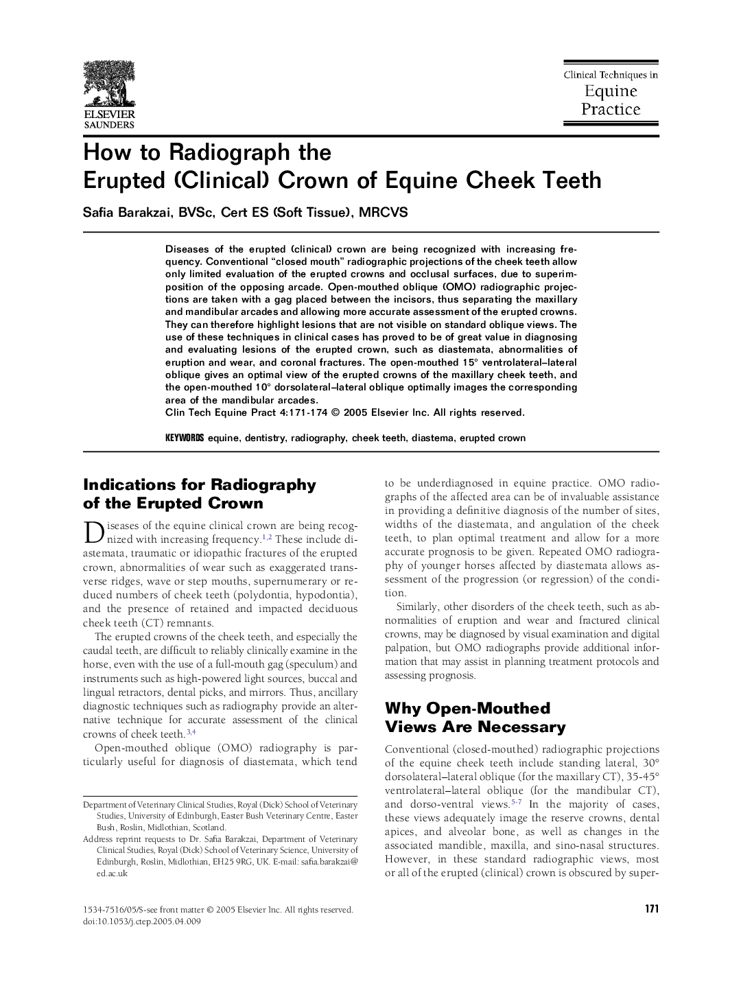
Diseases of the erupted (clinical) crown are being recognized with increasing frequency. Conventional “closed mouth” radiographic projections of the cheek teeth allow only limited evaluation of the erupted crowns and occlusal surfaces, due to superimposition of the opposing arcade. Open-mouthed oblique (OMO) radiographic projections are taken with a gag placed between the incisors, thus separating the maxillary and mandibular arcades and allowing more accurate assessment of the erupted crowns. They can therefore highlight lesions that are not visible on standard oblique views. The use of these techniques in clinical cases has proved to be of great value in diagnosing and evaluating lesions of the erupted crown, such as diastemata, abnormalities of eruption and wear, and coronal fractures. The open-mouthed 15° ventrolateral-lateral oblique gives an optimal view of the erupted crowns of the maxillary cheek teeth, and the open-mouthed 10° dorsolateral-lateral oblique optimally images the corresponding area of the mandibular arcades.
ناشر
Database: Elsevier - ScienceDirect (ساینس دایرکت)
Journal: Clinical Techniques in Equine Practice - Volume 4, Issue 2, June 2005, Pages 171-174
Journal: Clinical Techniques in Equine Practice - Volume 4, Issue 2, June 2005, Pages 171-174
نویسندگان
Safia BVSc, Cert ES (Soft Tissue), MRCVS,