| کد مقاله | کد نشریه | سال انتشار | مقاله انگلیسی | نسخه تمام متن |
|---|---|---|---|---|
| 9350220 | 1603714 | 2005 | 5 صفحه PDF | دانلود رایگان |
عنوان انگلیسی مقاله ISI
Le vissage percutané du scaphoïde assisté par ordinateur : étude expérimentale
دانلود مقاله + سفارش ترجمه
دانلود مقاله ISI انگلیسی
رایگان برای ایرانیان
کلمات کلیدی
موضوعات مرتبط
علوم پزشکی و سلامت
پزشکی و دندانپزشکی
ارتوپدی، پزشکی ورزشی و توانبخشی
پیش نمایش صفحه اول مقاله

چکیده انگلیسی
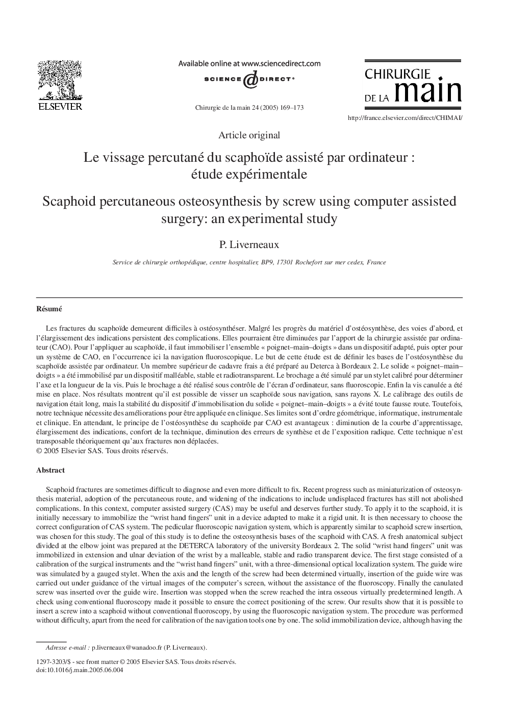
Scaphoid fractures are sometimes difficult to diagnose and even more difficult to fix. Recent progress such as miniaturization of osteosynthesis material, adoption of the percutaneous route, and widening of the indications to include undisplaced fractures has still not abolished complications. In this context, computer assisted surgery (CAS) may be useful and deserves further study. To apply it to the scaphoid, it is initially necessary to immobilize the “wrist hand fingers” unit in a device adapted to make it a rigid unit. It is then necessary to choose the correct configuration of CAS system. The pedicular fluoroscopic navigation system, which is apparently similar to scaphoid screw insertion, was chosen for this study. The goal of this study is to define the osteosynthesis bases of the scaphoid with CAS. A fresh anatomical subject divided at the elbow joint was prepared at the DETERCA laboratory of the university Bordeaux 2. The solid “wrist hand fingers” unit was immobilized in extension and ulnar deviation of the wrist by a malleable, stable and radio transparent device. The first stage consisted of a calibration of the surgical instruments and the “wrist hand fingers” unit, with a three-dimensional optical localization system. The guide wire was simulated by a gauged stylet. When the axis and the length of the screw had been determined virtually, insertion of the guide wire was carried out under guidance of the virtual images of the computer's screen, without the assistance of the fluoroscopy. Finally the canulated screw was inserted over the guide wire. Insertion was stopped when the screw reached the intra osseous virtually predetermined length. A check using conventional fluoroscopy made it possible to ensure the correct positioning of the screw. Our results show that it is possible to insert a screw into a scaphoid without conventional fluoroscopy, by using the fluoroscopic navigation system. The procedure was performed without difficulty, apart from the need for calibration of the navigation tools one by one. The solid immobilization device, although having the potential for micromovement, did not lead to any misdirection of the guide wire or screw. Our technique cannot be employed at the moment in live human surgery. Its limits are of a geometrical nature “two images available in two planes”, data processing “non-specific software dedicated”, instrumental “instrument calibration, micromobility of the immobilisation device” and live surgery “no current validation on a fractured scaphoid”. Meantime, the development of a percutaneous scaphoid osteosynthesis procedure by CAS can only bring advantages: reduction in the learning curve, widening of the indications, comfort in the technique, reduction in the errors of ostesynthesis and, reduction in the exposure to x-rays. In the current state of knowledge, the method would only be applicable to undisplaced fractures.
ناشر
Database: Elsevier - ScienceDirect (ساینس دایرکت)
Journal: Chirurgie de la Main - Volume 24, Issues 3â4, JuneâAugust 2005, Pages 169-173
Journal: Chirurgie de la Main - Volume 24, Issues 3â4, JuneâAugust 2005, Pages 169-173
نویسندگان
P. Liverneaux,