| کد مقاله | کد نشریه | سال انتشار | مقاله انگلیسی | نسخه تمام متن |
|---|---|---|---|---|
| 9356855 | 1267377 | 2005 | 13 صفحه PDF | دانلود رایگان |
عنوان انگلیسی مقاله ISI
Magnetic Resonance Arthrography of the Hip in Femoroacetabular Impingement: Technique and Findings
دانلود مقاله + سفارش ترجمه
دانلود مقاله ISI انگلیسی
رایگان برای ایرانیان
کلمات کلیدی
موضوعات مرتبط
علوم پزشکی و سلامت
پزشکی و دندانپزشکی
ارتوپدی، پزشکی ورزشی و توانبخشی
پیش نمایش صفحه اول مقاله

چکیده انگلیسی
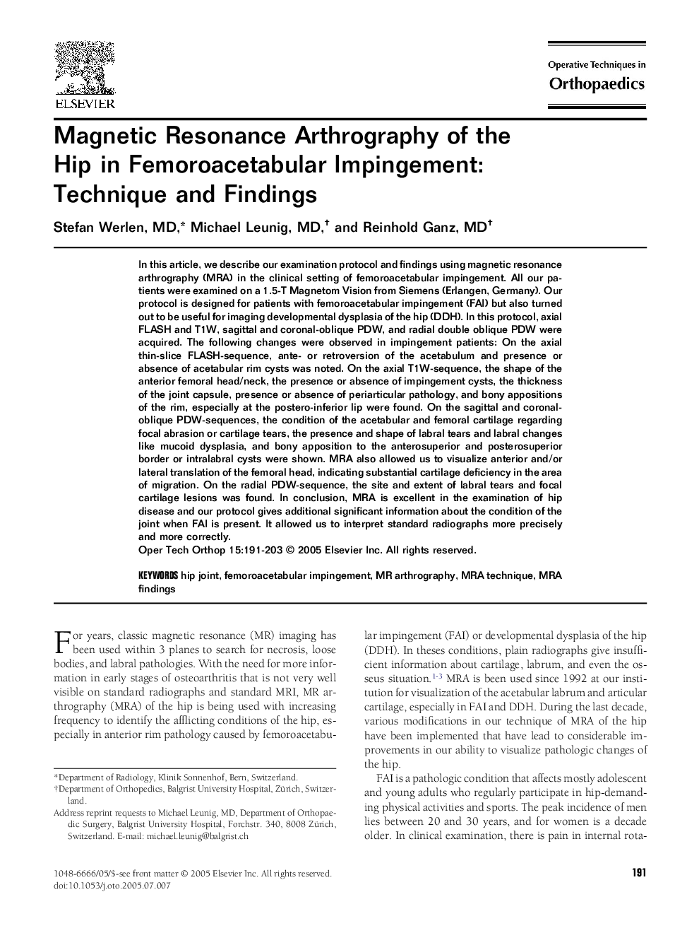
In this article, we describe our examination protocol and findings using magnetic resonance arthrography (MRA) in the clinical setting of femoroacetabular impingement. All our patients were examined on a 1.5-T Magnetom Vision from Siemens (Erlangen, Germany). Our protocol is designed for patients with femoroacetabular impingement (FAI) but also turned out to be useful for imaging developmental dysplasia of the hip (DDH). In this protocol, axial FLASH and T1W, sagittal and coronal-oblique PDW, and radial double oblique PDW were acquired. The following changes were observed in impingement patients: On the axial thin-slice FLASH-sequence, ante- or retroversion of the acetabulum and presence or absence of acetabular rim cysts was noted. On the axial T1W-sequence, the shape of the anterior femoral head/neck, the presence or absence of impingement cysts, the thickness of the joint capsule, presence or absence of periarticular pathology, and bony appositions of the rim, especially at the postero-inferior lip were found. On the sagittal and coronal-oblique PDW-sequences, the condition of the acetabular and femoral cartilage regarding focal abrasion or cartilage tears, the presence and shape of labral tears and labral changes like mucoid dysplasia, and bony apposition to the anterosuperior and posterosuperior border or intralabral cysts were shown. MRA also allowed us to visualize anterior and/or lateral translation of the femoral head, indicating substantial cartilage deficiency in the area of migration. On the radial PDW-sequence, the site and extent of labral tears and focal cartilage lesions was found. In conclusion, MRA is excellent in the examination of hip disease and our protocol gives additional significant information about the condition of the joint when FAI is present. It allowed us to interpret standard radiographs more precisely and more correctly.
ناشر
Database: Elsevier - ScienceDirect (ساینس دایرکت)
Journal: Operative Techniques in Orthopaedics - Volume 15, Issue 3, July 2005, Pages 191-203
Journal: Operative Techniques in Orthopaedics - Volume 15, Issue 3, July 2005, Pages 191-203
نویسندگان
Stefan MD, Michael MD, Reinhold MD,