| کد مقاله | کد نشریه | سال انتشار | مقاله انگلیسی | نسخه تمام متن |
|---|---|---|---|---|
| 9365411 | 1271506 | 2005 | 7 صفحه PDF | دانلود رایگان |
عنوان انگلیسی مقاله ISI
A review of 55 cases of cutaneous lymphoid hyperplasia: reassessment of the histopathologic findings leading to reclassification of 4 lesions as cutaneous marginal zone lymphoma and 19 as pseudolymphomatous folliculitis
دانلود مقاله + سفارش ترجمه
دانلود مقاله ISI انگلیسی
رایگان برای ایرانیان
کلمات کلیدی
PLFIgHCLHcutaneous pseudolymphomaImmunoglobulin heavy chain - زنجیره سنگین ایمونوگلوبولینDiffuse large B-cell lymphoma - لنفوم سلول B بزرگ سلول بزرگMarginal zone lymphoma - لنفوم ناحیه لگنCutaneous lymphoid hyperplasia - هیپرپلازی لنفوئیدی پوستیpolymerase chain reaction - واکنش زنجیره ای پلیمرازPCR - واکنش زنجیرهٔ پلیمراز
موضوعات مرتبط
علوم پزشکی و سلامت
پزشکی و دندانپزشکی
آسیبشناسی و فناوری پزشکی
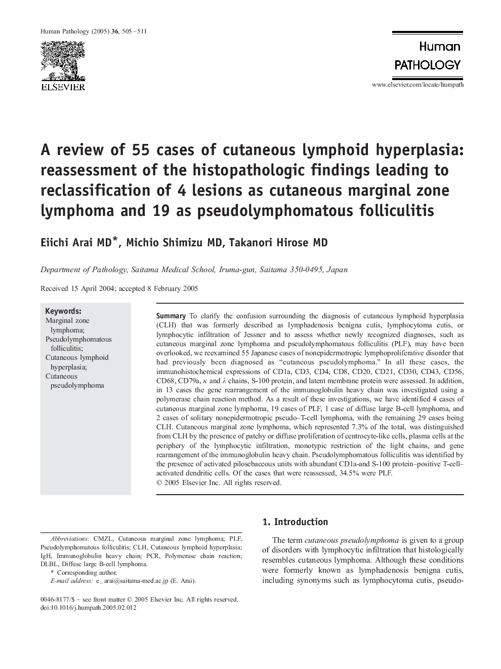
پیش نمایش صفحه اول مقاله

چکیده انگلیسی
To clarify the confusion surrounding the diagnosis of cutaneous lymphoid hyperplasia (CLH) that was formerly described as lymphadenosis benigna cutis, lymphocytoma cutis, or lymphocytic infiltration of Jessner and to assess whether newly recognized diagnoses, such as cutaneous marginal zone lymphoma and pseudolymphomatous folliculitis (PLF), may have been overlooked, we reexamined 55 Japanese cases of nonepidermotropic lymphoproliferative disorder that had previously been diagnosed as “cutaneous pseudolymphoma.” In all these cases, the immunohistochemical expressions of CD1a, CD3, CD4, CD8, CD20, CD21, CD30, CD43, CD56, CD68, CD79a, κ and λ chains, S-100 protein, and latent membrane protein were assessed. In addition, in 13 cases the gene rearrangement of the immunoglobulin heavy chain was investigated using a polymerase chain reaction method. As a result of these investigations, we have identified 4 cases of cutaneous marginal zone lymphoma, 19 cases of PLF, 1 case of diffuse large B-cell lymphoma, and 2 cases of solitary nonepidermotropic pseudo-T-cell lymphoma, with the remaining 29 cases being CLH. Cutaneous marginal zone lymphoma, which represented 7.3% of the total, was distinguished from CLH by the presence of patchy or diffuse proliferation of centrocyte-like cells, plasma cells at the periphery of the lymphocytic infiltration, monotypic restriction of the light chains, and gene rearrangement of the immunoglobulin heavy chain. Pseudolymphomatous folliculitis was identified by the presence of activated pilosebaceous units with abundant CD1a-and S-100 protein-positive T-cell-activated dendritic cells. Of the cases that were reassessed, 34.5% were PLF.
ناشر
Database: Elsevier - ScienceDirect (ساینس دایرکت)
Journal: Human Pathology - Volume 36, Issue 5, May 2005, Pages 505-511
Journal: Human Pathology - Volume 36, Issue 5, May 2005, Pages 505-511
نویسندگان
Eiichi MD, Michio MD, Takanori MD,