| کد مقاله | کد نشریه | سال انتشار | مقاله انگلیسی | نسخه تمام متن |
|---|---|---|---|---|
| 1229929 | 1495242 | 2014 | 9 صفحه PDF | دانلود رایگان |

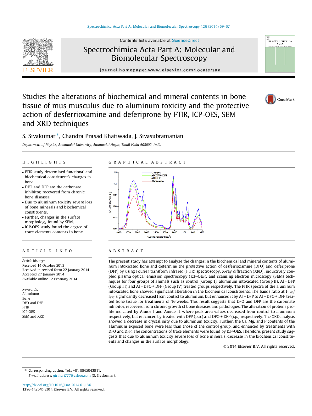
• FTIR study determined functional and biochemical constituent’s changes in bone.
• DFO and DFP are the carbonate inhibitor, recovered from chronic bone diseases.
• Due to aluminum toxicity severe loss of bone minerals and biochemical constituents.
• Further, changes in the surface morphology found by SEM.
• ICP-OES study found the degree of trace elements contents in bone.
The present study has attempt to analyze the changes in the biochemical and mineral contents of aluminum intoxicated bone and determine the protective action of desferrioxamine (DFO) and deferiprone (DFP) by using Fourier transform infrared (FTIR) spectroscopy, X-ray diffraction (XRD), inductively coupled plasma optical emission spectroscopy (ICP-OES), and scanning electron microscopy (SEM) techniques for four groups of animals such as control (Group I), aluminum intoxicated (Group II), Al + DFP (Group III) and Al + DFO + DFP (Group IV) treated groups respectively. The FTIR spectra of the aluminum intoxicated bone showed significant alteration in the biochemical constituents. The bands ratio at I1400/I877 significantly decreased from control to aluminum, but enhanced it by Al + DFP to Al + DFO + DFP treated bone tissue for treatments of 16 weeks. This result suggests that DFO and DFP are the carbonate inhibitor, recovered from chronic growth of bone diseases and pathologies. The alteration of proteins profile indicated by Amide I and Amide II, where peak area values decreased from control to aluminum respectively, but enhanced by treated with DFP (p.o.) and DFO + DFP (i.p.) respectively. The XRD analysis showed a decrease in crystallinity due to aluminum toxicity. Further, the Ca, Mg, and P contents of the aluminum exposed bone were less than those of the control group, and enhanced by treatments with DFO and DFP. The concentrations of trace elements were found by ICP-OES. Therefore, present study suggests that due to aluminum toxicity severe loss of bone minerals, decrease in the biochemical constituents and changes in the surface morphology.
Figure optionsDownload as PowerPoint slide
Journal: Spectrochimica Acta Part A: Molecular and Biomolecular Spectroscopy - Volume 126, 21 May 2014, Pages 59–67