| کد مقاله | کد نشریه | سال انتشار | مقاله انگلیسی | نسخه تمام متن |
|---|---|---|---|---|
| 1231643 | 1495219 | 2015 | 9 صفحه PDF | دانلود رایگان |
• The ultrastructure on the interaction of shikonin and HIgG was visually displayed.
• The present of shinkonin induced the change of conformation and secondary structure of HIgG.
• The binding and thermodynamic parameters illustrated the acting mechanism of shinkonin and HIgG at molecule level.
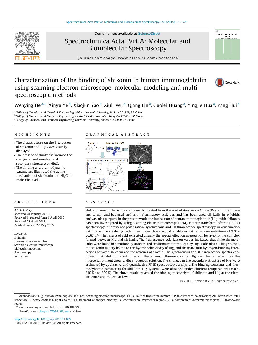
Shikonin, one of the active components isolated from the root of Arnebia euchroma (Royle) Johnst, have anti-tumor, anti-bacterial and anti-inflammatory activities and has been used clinically in phlebitis and vascular purpura. In the present work, the interaction of human immunoglobulin (HIg) with shikonin has been investigated by using scanning electron microscope (SEM), Fourier transform infrared (FT-IR) spectroscopy, fluorescence polarization, synchronous and 3D fluorescence spectroscopy in combination with molecular modeling techniques under physiological conditions with drug concentrations of 3.33–36.67 μM. The results of SEM exhibited visually the special effect on aggregation behavior of the complex formed between HIg and shikonin. The fluorescence polarization values indicated that shikonin molecules were found in a motionally unrestricted environment introduced by HIg. Molecular docking showed the shikonin moiety bound to the hydrophobic cavity of HIg, and there are four hydrogen-bonding interactions between shikonin and the residues of protein. The synchronous and 3D fluorescence spectra confirmed that shikonin could quench the intrinsic fluorescence of HIg and has an effect on the microenvironment around HIg in aqueous solution. The changes in the secondary structure of HIg were estimated by qualitative and quantitative FT-IR spectroscopic analysis. The binding constants and thermodynamic parameters for shikonin–HIg systems were obtained under different temperatures (300 K, 310 K and 320 K). The above results revealed the binding mechanism of shikonin and HIg at the ultrastructure and molecular level.
Figure optionsDownload as PowerPoint slide
Journal: Spectrochimica Acta Part A: Molecular and Biomolecular Spectroscopy - Volume 150, 5 November 2015, Pages 514–522
