| کد مقاله | کد نشریه | سال انتشار | مقاله انگلیسی | نسخه تمام متن |
|---|---|---|---|---|
| 1850107 | 1528247 | 2006 | 7 صفحه PDF | دانلود رایگان |
عنوان انگلیسی مقاله ISI
2D and 3D refraction-based visualization of breast cancer for early clinical check
دانلود مقاله + سفارش ترجمه
دانلود مقاله ISI انگلیسی
رایگان برای ایرانیان
کلمات کلیدی
موضوعات مرتبط
مهندسی و علوم پایه
فیزیک و نجوم
فیزیک هسته ای و انرژی بالا
پیش نمایش صفحه اول مقاله

چکیده انگلیسی
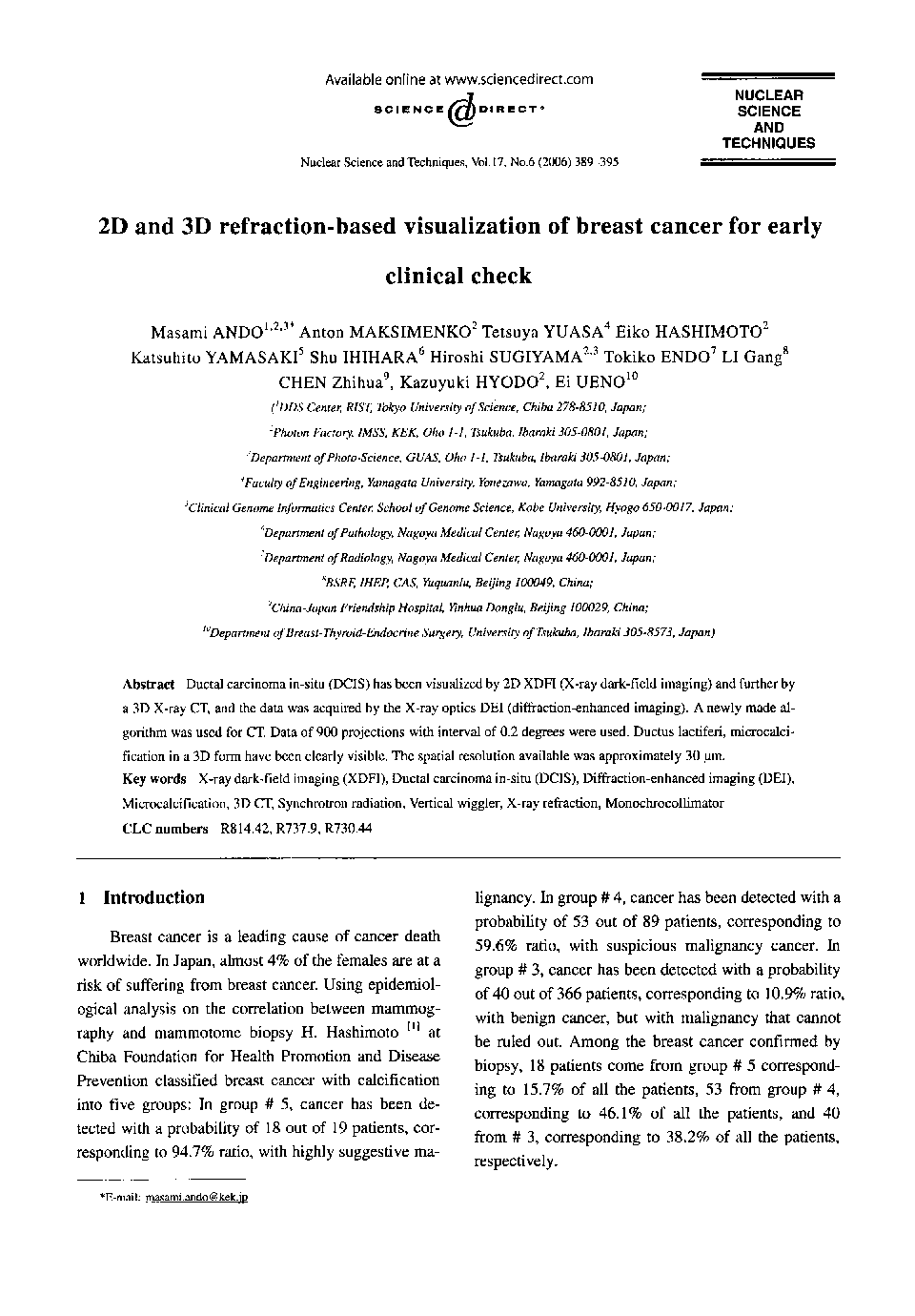
Ductal carcinoma in-situ (DCIS) has been visualized by 2D XDFI (X-ray dark-field imaging) and further by a 3D X-ray CT, and the data was acquired by the X-ray optics DEI (diffraction-enhanced imaging). A newly made algorithm was used for CT. Data of 900 projections with interval of 0.2 degrees were used. Ductus lactiferi, microcalcification in a 3D form have been clearly visible. The spatial resolution available was approximately 30 μm.
ناشر
Database: Elsevier - ScienceDirect (ساینس دایرکت)
Journal: Nuclear Science and Techniques - Volume 17, Issue 6, December 2006, Pages 389-395
Journal: Nuclear Science and Techniques - Volume 17, Issue 6, December 2006, Pages 389-395
نویسندگان
Masami ANDO, Anton MAKSIMENKO, Tetsuya YUASA, Eiko HASHIMOTO, Katsuhito YAMASAKI, Shu IHIHARA, Hiroshi SUGIYAMA, Tokiko ENDO, LI Gang, CHEN Zhihua, Kazuyuki HYODO, Ei UENO,