| کد مقاله | کد نشریه | سال انتشار | مقاله انگلیسی | نسخه تمام متن |
|---|---|---|---|---|
| 2019005 | 1068378 | 2013 | 123 صفحه PDF | دانلود رایگان |
عنوان انگلیسی مقاله ISI
Image Cytometry: Protocols for 2D and 3D Quantification in Microscopic Images
دانلود مقاله + سفارش ترجمه
دانلود مقاله ISI انگلیسی
رایگان برای ایرانیان
موضوعات مرتبط
علوم زیستی و بیوفناوری
بیوشیمی، ژنتیک و زیست شناسی مولکولی
زیست شیمی
پیش نمایش صفحه اول مقاله

چکیده انگلیسی
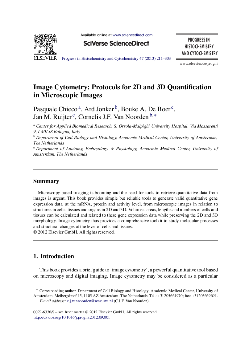
SummaryMicroscopy-based imaging is booming and the need for tools to retrieve quantitative data from images is urgent. This book provides simple but reliable tools to generate valid quantitative gene expression data, at the mRNA, protein and activity level, from microscopic images in relation to structures in cells, tissues and organs in 2D and 3D. Volumes, areas, lengths and numbers of cells and tissues can be calculated and related to these gene expression data while preserving the 2D and 3D morphology. Image cytometry thus provides a comprehensive toolkit to study molecular processes and structural changes at the level of cells and tissues.
ناشر
Database: Elsevier - ScienceDirect (ساینس دایرکت)
Journal: Progress in Histochemistry and Cytochemistry - Volume 47, Issue 4, January 2013, Pages 211–333
Journal: Progress in Histochemistry and Cytochemistry - Volume 47, Issue 4, January 2013, Pages 211–333
نویسندگان
Pasquale Chieco, Ard Jonker, Bouke A. De Boer, Jan M. Ruijter, Cornelis J.F. Van Noorden,