| کد مقاله | کد نشریه | سال انتشار | مقاله انگلیسی | نسخه تمام متن |
|---|---|---|---|---|
| 2769599 | 1151320 | 2008 | 10 صفحه PDF | دانلود رایگان |
عنوان انگلیسی مقاله ISI
EcografÃa portátil en anestesia regional: bloqueos del plexo braquial
دانلود مقاله + سفارش ترجمه
دانلود مقاله ISI انگلیسی
رایگان برای ایرانیان
کلمات کلیدی
موضوعات مرتبط
علوم پزشکی و سلامت
پزشکی و دندانپزشکی
بیهوشی و پزشکی درد
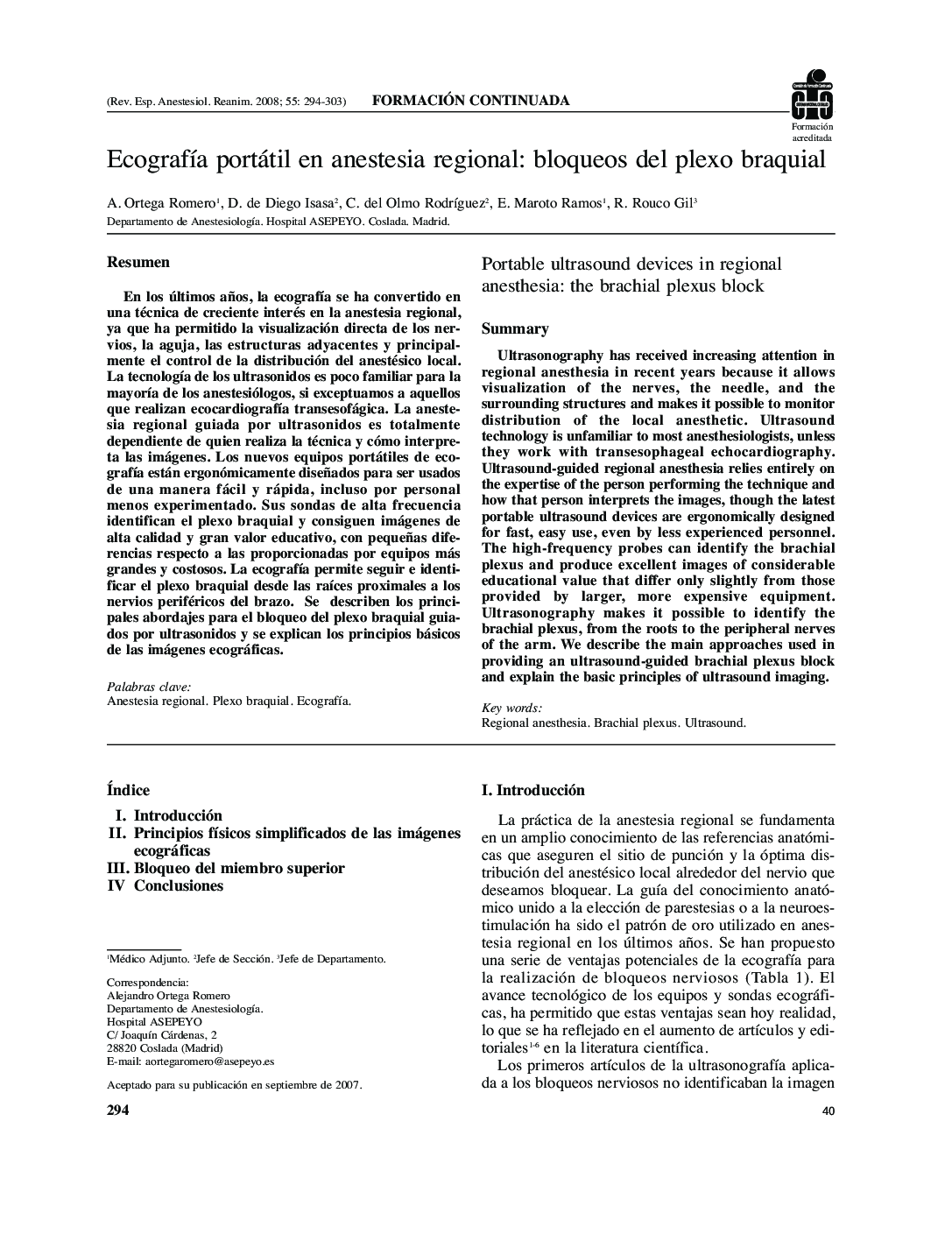
پیش نمایش صفحه اول مقاله

چکیده انگلیسی
Ultrasonography has received increasing attention in regional anesthesia in recent years because it allows visualization of the nerves, the needle, and the surrounding structures and makes it possible to monitor distribution of the local anesthetic. Ultrasound technology is unfamiliar to most anesthesiologists, unless they work with transesophageal echocardiography. Ultrasound-guided regional anesthesia relies entirely on the expertise of the person performing the technique and how that person interprets the images, though the latest portable ultrasound devices are ergonomically designed for fast, easy use, even by less experienced personnel. The high-frequency probes can identify the brachial plexus and produce excellent images of considerable educational value that differ only slightly from those provided by larger, more expensive equipment. Ultrasonography makes it possible to identify the brachial plexus, from the roots to the peripheral nerves of the arm. We describe the main approaches used in providing an ultrasound-guided brachial plexus block and explain the basic principles of ultrasound imaging.
ناشر
Database: Elsevier - ScienceDirect (ساینس دایرکت)
Journal: Revista Española de AnestesiologÃa y Reanimación - Volume 55, Issue 5, May 2008, Pages 294-303
Journal: Revista Española de AnestesiologÃa y Reanimación - Volume 55, Issue 5, May 2008, Pages 294-303
نویسندگان
A. (Médico Adjunto), D. (Jefe de Sección), C. (Jefe de Sección), E. (Médico Adjunto), R. (Jefe de Departamento),