| کد مقاله | کد نشریه | سال انتشار | مقاله انگلیسی | نسخه تمام متن |
|---|---|---|---|---|
| 2840097 | 1165244 | 2007 | 7 صفحه PDF | دانلود رایگان |
عنوان انگلیسی مقاله ISI
TomografÃa computarizada multidetectora en el estudio cardÃaco
دانلود مقاله + سفارش ترجمه
دانلود مقاله ISI انگلیسی
رایگان برای ایرانیان
کلمات کلیدی
موضوعات مرتبط
علوم زیستی و بیوفناوری
بیوشیمی، ژنتیک و زیست شناسی مولکولی
فیزیولوژی
پیش نمایش صفحه اول مقاله

چکیده انگلیسی
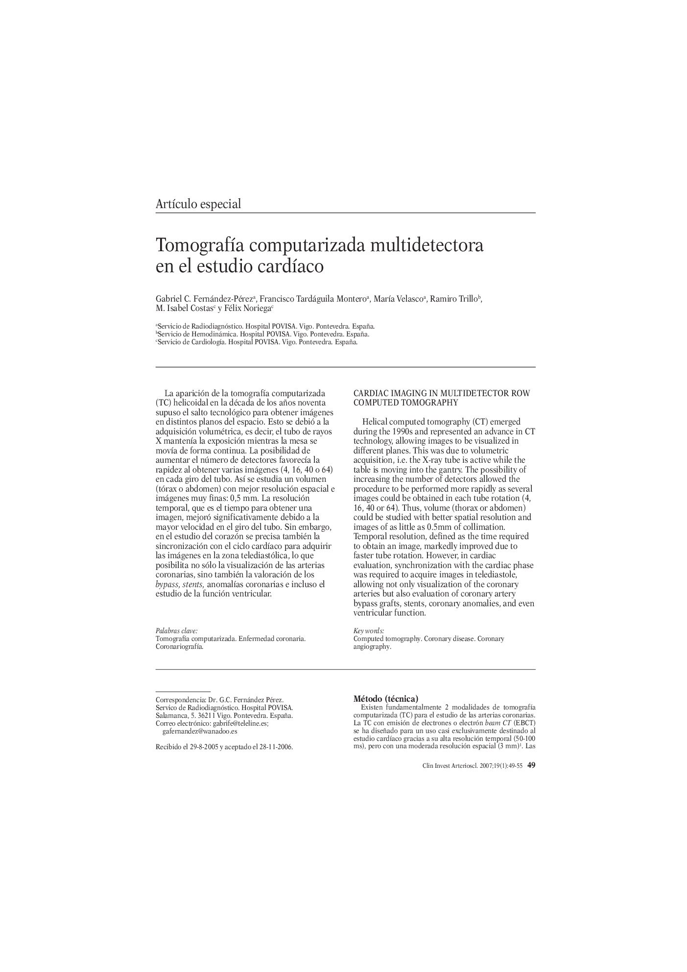
Helical computed tomography (CT) emerged during the 1990s and represented an advance in CT technology, allowing images to be visualized in different planes. This was due to volumetric acquisition, i.e. the X-ray tube is active while the table is moving into the gantry. The possibility of increasing the number of detectors allowed the procedure to be performed more rapidly as several images could be obtained in each tube rotation (4, 16, 40 or 64). Thus, volume (thorax or abdomen) could be studied with better spatial resolution and images of as little as 0.5mm of collimation. Temporal resolution, defined as the time required to obtain an image, markedly improved due to faster tube rotation. However, in cardiac evaluation, synchronization with the cardiac phase was required to acquire images in telediastole, allowing not only visualization of the coronary arteries but also evaluation of coronary artery bypass grafts, stents, coronary anomalies, and even ventricular function.
ناشر
Database: Elsevier - ScienceDirect (ساینس دایرکت)
Journal: ClÃnica e Investigación en Arteriosclerosis - Volume 19, Issue 1, February 2007, Pages 49-55
Journal: ClÃnica e Investigación en Arteriosclerosis - Volume 19, Issue 1, February 2007, Pages 49-55
نویسندگان
Gabriel C. Fernández-Pérez, Francisco Tardáguila Montero, MarÃa Velasco, Ramiro Trillo, M. Isabel Costas, Félix Noriega,