| کد مقاله | کد نشریه | سال انتشار | مقاله انگلیسی | نسخه تمام متن |
|---|---|---|---|---|
| 3334638 | 1213455 | 2009 | 22 صفحه PDF | دانلود رایگان |
عنوان انگلیسی مقاله ISI
Lesions of the Nipple
دانلود مقاله + سفارش ترجمه
دانلود مقاله ISI انگلیسی
رایگان برای ایرانیان
موضوعات مرتبط
علوم پزشکی و سلامت
پزشکی و دندانپزشکی
هماتولوژی
پیش نمایش صفحه اول مقاله

چکیده انگلیسی
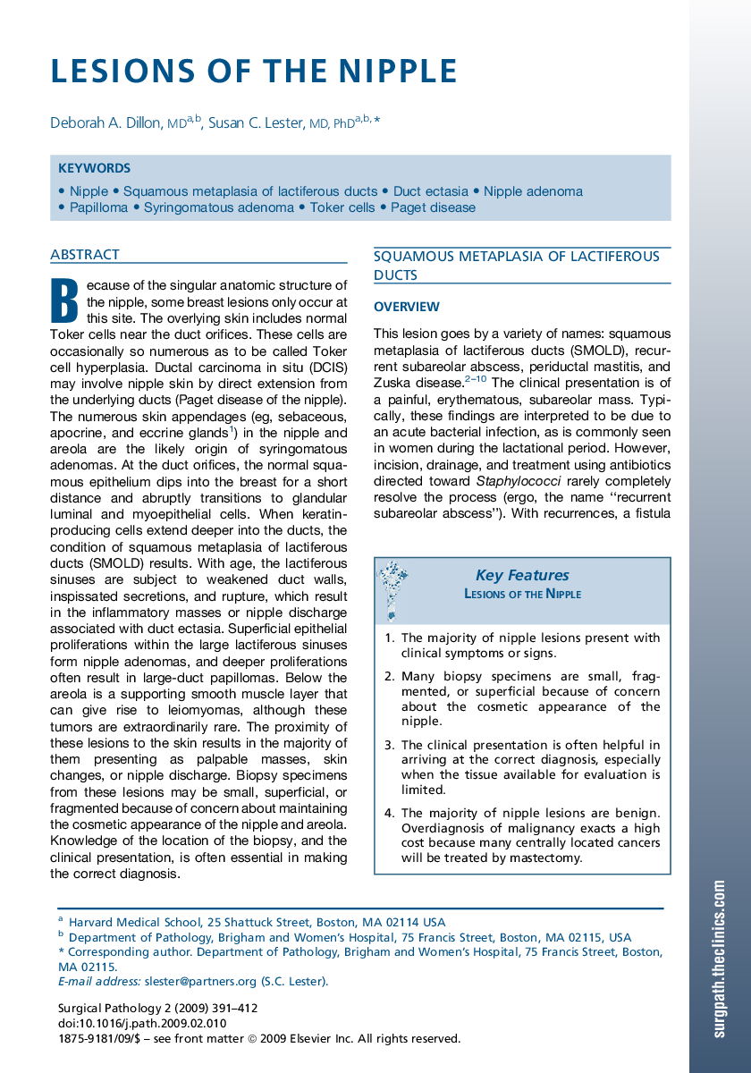
Because of the singular anatomic structure of the nipple, some breast lesions only occur at this site. The overlying skin includes normal Toker cells near the duct orifices. These cells are occasionally so numerous as to be called Toker cell hyperplasia. Ductal carcinoma in situ (DCIS) may involve nipple skin by direct extension from the underlying ducts (Paget disease of the nipple). The numerous skin appendages (eg, sebaceous, apocrine, and eccrine glands1) in the nipple and areola are the likely origin of syringomatous adenomas. At the duct orifices, the normal squamous epithelium dips into the breast for a short distance and abruptly transitions to glandular luminal and myoepithelial cells. When keratin-producing cells extend deeper into the ducts, the condition of squamous metaplasia of lactiferous ducts (SMOLD) results. With age, the lactiferous sinuses are subject to weakened duct walls, inspissated secretions, and rupture, which result in the inflammatory masses or nipple discharge associated with duct ectasia. Superficial epithelial proliferations within the large lactiferous sinuses form nipple adenomas, and deeper proliferations often result in large-duct papillomas. Below the areola is a supporting smooth muscle layer that can give rise to leiomyomas, although these tumors are extraordinarily rare. The proximity of these lesions to the skin results in the majority of them presenting as palpable masses, skin changes, or nipple discharge. Biopsy specimens from these lesions may be small, superficial, or fragmented because of concern about maintaining the cosmetic appearance of the nipple and areola. Knowledge of the location of the biopsy, and the clinical presentation, is often essential in making the correct diagnosis.Key FeaturesLesions of the Nipple1.The majority of nipple lesions present with clinical symptoms or signs.2.Many biopsy specimens are small, fragmented, or superficial because of concern about the cosmetic appearance of the nipple.3.The clinical presentation is often helpful in arriving at the correct diagnosis, especially when the tissue available for evaluation is limited.4.The majority of nipple lesions are benign. Overdiagnosis of malignancy exacts a high cost because many centrally located cancers will be treated by mastectomy.
ناشر
Database: Elsevier - ScienceDirect (ساینس دایرکت)
Journal: Surgical Pathology Clinics - Volume 2, Issue 2, June 2009, Pages 391-412
Journal: Surgical Pathology Clinics - Volume 2, Issue 2, June 2009, Pages 391-412
نویسندگان
Deborah A. MD, Susan C. MD, PhD,