| کد مقاله | کد نشریه | سال انتشار | مقاله انگلیسی | نسخه تمام متن |
|---|---|---|---|---|
| 3469002 | 1232733 | 2014 | 17 صفحه PDF | دانلود رایگان |
عنوان انگلیسی مقاله ISI
Apport du scanner dans la pathologie du carrefour iléo-cæcal
دانلود مقاله + سفارش ترجمه
دانلود مقاله ISI انگلیسی
رایگان برای ایرانیان
کلمات کلیدی
adénocarcinome - آدنوکارسینومAdenocarcinoma - آدنوکارسینوماAppendicite - آپاندیسیتAppendicitis - آپاندیسیت یا آویزآماسScanner - اسکنرIschemia - ایسکمیIschémie - ایسکمیIleitis - ایلیتTuberculosis - بیماری سلCrohn's disease - بیماری کرونLymphoma - لنفومLymphome - لنفومMaladie de Crohn - مالدی د کرونTuberculose - مرض سلCarcinoïde - کارسینوئیدcarcinoid - کارسینوئید، تومور کارسینوئید
موضوعات مرتبط
علوم پزشکی و سلامت
پزشکی و دندانپزشکی
پزشکی و دندانپزشکی (عمومی)
پیش نمایش صفحه اول مقاله

چکیده انگلیسی
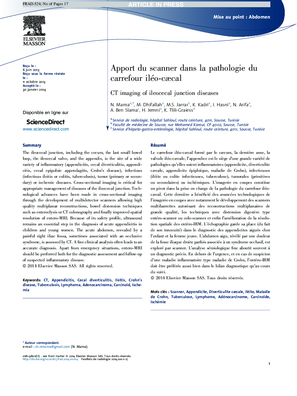
The ileocecal junction, including the cecum, the last small bowel loop, the ileocecal valve, and the appendix, is the site of a wide variety of inflammatory (appendicitis, cecal diverticulitis, appendicitis, cecal epipoloic appendagitis, Crohn's disease), infectious (infectious ileitis or colitis, tuberculosis), tumor (primary or secondary) or ischemic diseases. Cross-sectional imaging is critical for appropriate management of diseases of the ileocecal junction. Technological advances have been made in cross-sectional imaging through the development of multidetector scanners allowing high quality multiplanar reconstructions, bowel distension techniques such as enteroclysis or CT colonography and finally improved spatial resolution of entero-MRI. Because of its safety profile, ultrasound remains an essential step in the diagnosis of acute appendicitis in children and young women. The acute abdomen, revealed by a painful right iliac fossa, sometimes associated with an occlusive syndrome, is assessed by CT. A fine clinical analysis often leads to an accurate diagnosis. Apart from emergency situations, entero-MRI should be preferred both for the diagnostic assessment and follow-up of suspected inflammatory disease.
ناشر
Database: Elsevier - ScienceDirect (ساینس دایرکت)
Journal: Feuillets de Radiologie - Volume 54, Issue 5, October 2014, Pages 275-291
Journal: Feuillets de Radiologie - Volume 54, Issue 5, October 2014, Pages 275-291
نویسندگان
N. Mama, M. Dhifallah, M.S. Jarrar, K. Kadri, I. Hasni, N. Arifa, A. Ben Slama, H. Jemni, K. Tlili-Graiess,