| کد مقاله | کد نشریه | سال انتشار | مقاله انگلیسی | نسخه تمام متن |
|---|---|---|---|---|
| 3841414 | 1247978 | 2008 | 9 صفحه PDF | دانلود رایگان |
عنوان انگلیسی مقاله ISI
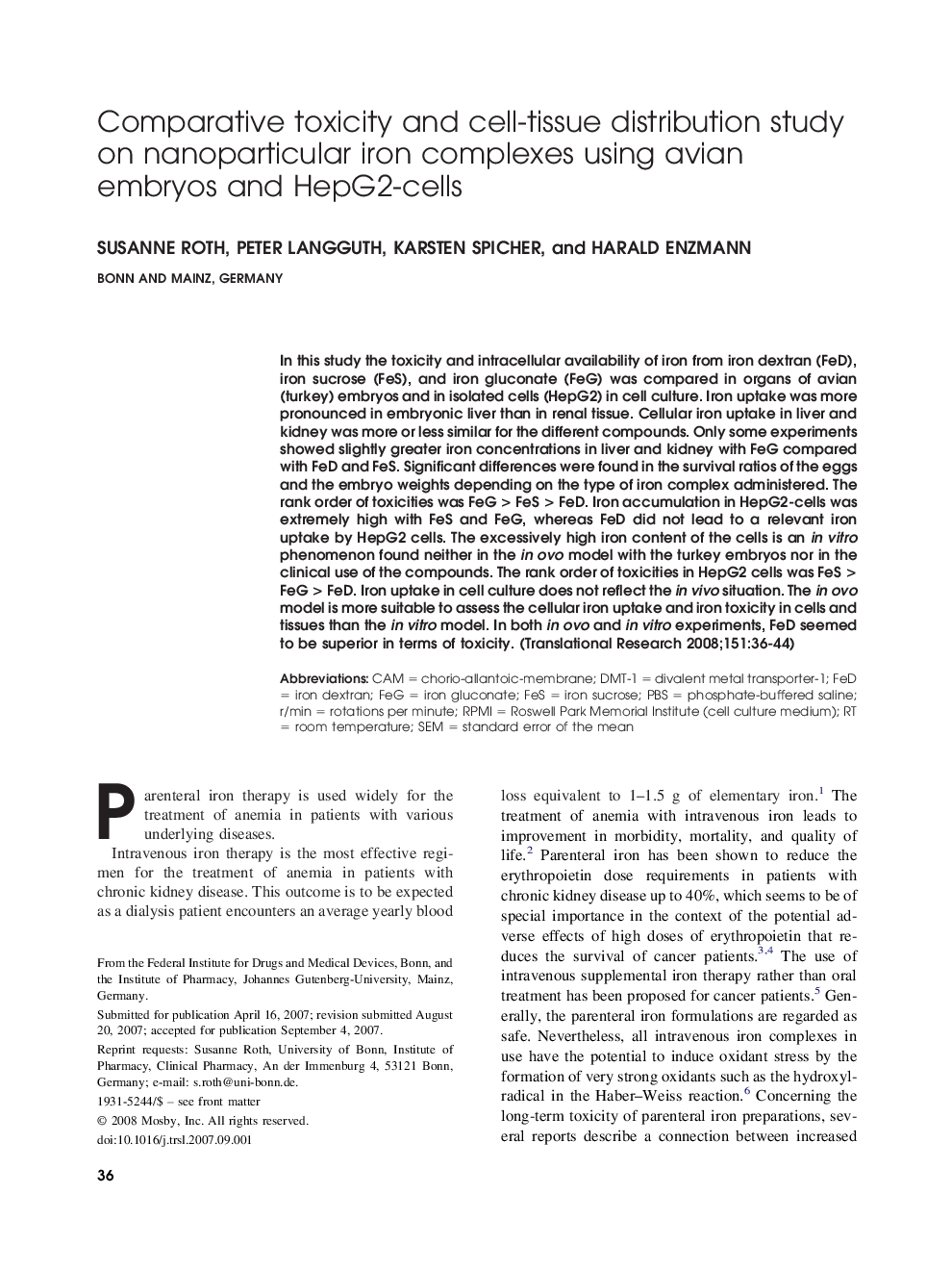
Comparative toxicity and cell-tissue distribution study on nanoparticular iron complexes using avian embryos and HepG2-cells
دانلود مقاله + سفارش ترجمه
دانلود مقاله ISI انگلیسی
رایگان برای ایرانیان
کلمات کلیدی
PBSFeSRPMIDMT-1standard error of the mean - خطای استاندارد میانگینRoom temperature - دمای اتاقIron dextran - دکتران آهنCAM - ساخت به کمک کامپیوترIron sucrose - ساکارز آهنFed - فدرال رزروdivalent metal transporter-1 - فلات دوالانت فلزی 1Phosphate-buffered saline - محلول نمک فسفات با خاصیت بافریSEM - مدل معادلات ساختاری / میکروسکوپ الکترونی روبشیfeG - مرغrotations per minute - چرخش در دقیقهiron gluconate - گلوکونات آهن
موضوعات مرتبط
علوم پزشکی و سلامت
پزشکی و دندانپزشکی
پزشکی و دندانپزشکی (عمومی)
پیش نمایش صفحه اول مقاله

چکیده انگلیسی
In this study the toxicity and intracellular availability of iron from iron dextran (FeD), iron sucrose (FeS), and iron gluconate (FeG) was compared in organs of avian (turkey) embryos and in isolated cells (HepG2) in cell culture. Iron uptake was more pronounced in embryonic liver than in renal tissue. Cellular iron uptake in liver and kidney was more or less similar for the different compounds. Only some experiments showed slightly greater iron concentrations in liver and kidney with FeG compared with FeD and FeS. Significant differences were found in the survival ratios of the eggs and the embryo weights depending on the type of iron complex administered. The rank order of toxicities was FeG > FeS > FeD. Iron accumulation in HepG2-cells was extremely high with FeS and FeG, whereas FeD did not lead to a relevant iron uptake by HepG2 cells. The excessively high iron content of the cells is an in vitro phenomenon found neither in the in ovo model with the turkey embryos nor in the clinical use of the compounds. The rank order of toxicities in HepG2 cells was FeS > FeG > FeD. Iron uptake in cell culture does not reflect the in vivo situation. The in ovo model is more suitable to assess the cellular iron uptake and iron toxicity in cells and tissues than the in vitro model. In both in ovo and in vitro experiments, FeD seemed to be superior in terms of toxicity.
ناشر
Database: Elsevier - ScienceDirect (ساینس دایرکت)
Journal: Translational Research - Volume 151, Issue 1, January 2008, Pages 36-44
Journal: Translational Research - Volume 151, Issue 1, January 2008, Pages 36-44
نویسندگان
Susanne Roth, Peter Langguth, Karsten Spicher, Harald Enzmann,