| کد مقاله | کد نشریه | سال انتشار | مقاله انگلیسی | نسخه تمام متن |
|---|---|---|---|---|
| 4095111 | 1268512 | 2007 | 7 صفحه PDF | دانلود رایگان |
عنوان انگلیسی مقاله ISI
Current Status of Imaging of the Intervertebral Disc
دانلود مقاله + سفارش ترجمه
دانلود مقاله ISI انگلیسی
رایگان برای ایرانیان
کلمات کلیدی
موضوعات مرتبط
علوم پزشکی و سلامت
پزشکی و دندانپزشکی
ارتوپدی، پزشکی ورزشی و توانبخشی
پیش نمایش صفحه اول مقاله

چکیده انگلیسی
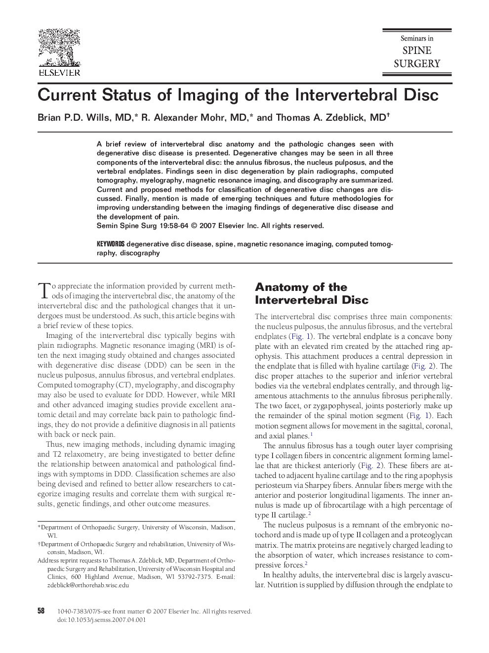
A brief review of intervertebral disc anatomy and the pathologic changes seen with degenerative disc disease is presented. Degenerative changes may be seen in all three components of the intervertebral disc: the annulus fibrosus, the nucleus pulposus, and the vertebral endplates. Findings seen in disc degeneration by plain radiographs, computed tomography, myelography, magnetic resonance imaging, and discography are summarized. Current and proposed methods for classification of degenerative disc changes are discussed. Finally, mention is made of emerging techniques and future methodologies for improving understanding between the imaging findings of degenerative disc disease and the development of pain.
ناشر
Database: Elsevier - ScienceDirect (ساینس دایرکت)
Journal: Seminars in Spine Surgery - Volume 19, Issue 2, June 2007, Pages 58–64
Journal: Seminars in Spine Surgery - Volume 19, Issue 2, June 2007, Pages 58–64
نویسندگان
Brian P.D. Wills, R. Alexander Mohr, Thomas A. Zdeblick,