| کد مقاله | کد نشریه | سال انتشار | مقاله انگلیسی | نسخه تمام متن |
|---|---|---|---|---|
| 4337399 | 1614759 | 2016 | 13 صفحه PDF | دانلود رایگان |
• Expression of cerebral monocarboxylate transporters was determined 1 and 24 h following transient ischemia in mice.
• After 1 h, MCT1, MCT2 and MCT4 expressions were upregulated in the ipsilateral and contralateral striatum and cortex.
• Neuronal MCT2 but also MCT1 expression, astrocytic MCT4 expression and microvessel MCT1 expression all increased at 1 h.
• After 24 h, overall MCT2 and MCT4 expressions decreased in the ipsilateral and contralateral striatum and cortex.
• Neuronal MCT2 expression was reduced but MCT4 expression appeared while microvessels exhibited MCT2 expression at 24 h.
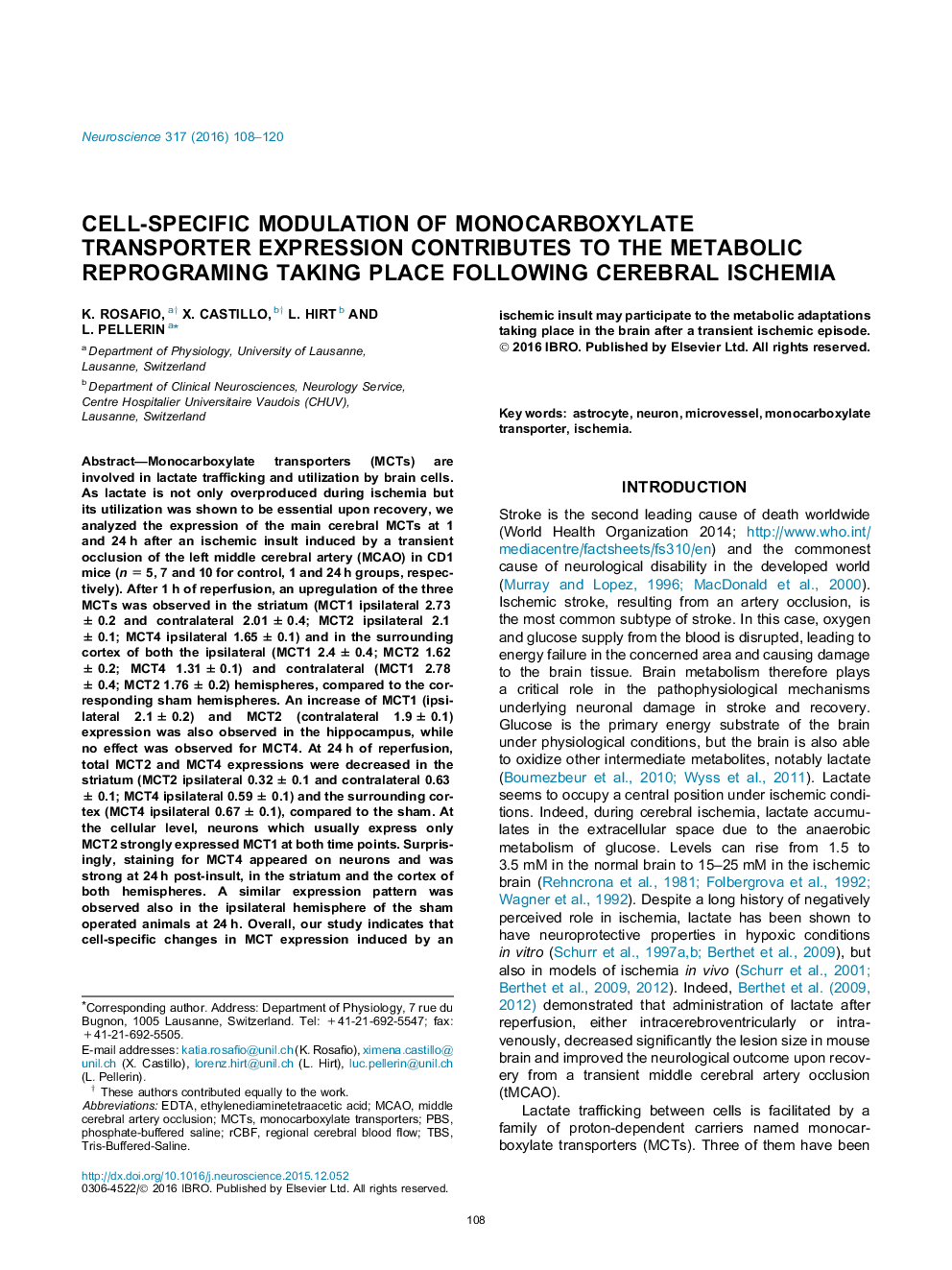
Monocarboxylate transporters (MCTs) are involved in lactate trafficking and utilization by brain cells. As lactate is not only overproduced during ischemia but its utilization was shown to be essential upon recovery, we analyzed the expression of the main cerebral MCTs at 1 and 24 h after an ischemic insult induced by a transient occlusion of the left middle cerebral artery (MCAO) in CD1 mice (n = 5, 7 and 10 for control, 1 and 24 h groups, respectively). After 1 h of reperfusion, an upregulation of the three MCTs was observed in the striatum (MCT1 ipsilateral 2.73 ± 0.2 and contralateral 2.01 ± 0.4; MCT2 ipsilateral 2.1 ± 0.1; MCT4 ipsilateral 1.65 ± 0.1) and in the surrounding cortex of both the ipsilateral (MCT1 2.4 ± 0.4; MCT2 1.62 ± 0.2; MCT4 1.31 ± 0.1) and contralateral (MCT1 2.78 ± 0.4; MCT2 1.76 ± 0.2) hemispheres, compared to the corresponding sham hemispheres. An increase of MCT1 (ipsilateral 2.1 ± 0.2) and MCT2 (contralateral 1.9 ± 0.1) expression was also observed in the hippocampus, while no effect was observed for MCT4. At 24 h of reperfusion, total MCT2 and MCT4 expressions were decreased in the striatum (MCT2 ipsilateral 0.32 ± 0.1 and contralateral 0.63 ± 0.1; MCT4 ipsilateral 0.59 ± 0.1) and the surrounding cortex (MCT4 ipsilateral 0.67 ± 0.1), compared to the sham. At the cellular level, neurons which usually express only MCT2 strongly expressed MCT1 at both time points. Surprisingly, staining for MCT4 appeared on neurons and was strong at 24 h post-insult, in the striatum and the cortex of both hemispheres. A similar expression pattern was observed also in the ipsilateral hemisphere of the sham operated animals at 24 h. Overall, our study indicates that cell-specific changes in MCT expression induced by an ischemic insult may participate to the metabolic adaptations taking place in the brain after a transient ischemic episode.
Figure optionsDownload high-quality image (129 K)Download as PowerPoint slide
Journal: Neuroscience - Volume 317, 11 March 2016, Pages 108–120
