| کد مقاله | کد نشریه | سال انتشار | مقاله انگلیسی | نسخه تمام متن |
|---|---|---|---|---|
| 5938576 | 1573465 | 2008 | 14 صفحه PDF | دانلود رایگان |
عنوان انگلیسی مقاله ISI
Longitudinal in Vivo Positron Emission Tomography Imaging of Infected and Activated Brain Macrophages in a Macaque Model of Human Immunodeficiency Virus Encephalitis Correlates with Central and Peripheral Markers of Encephalitis and Areas of Synaptic Dege
دانلود مقاله + سفارش ترجمه
دانلود مقاله ISI انگلیسی
رایگان برای ایرانیان
موضوعات مرتبط
علوم پزشکی و سلامت
پزشکی و دندانپزشکی
کاردیولوژی و پزشکی قلب و عروق
پیش نمایش صفحه اول مقاله

چکیده انگلیسی
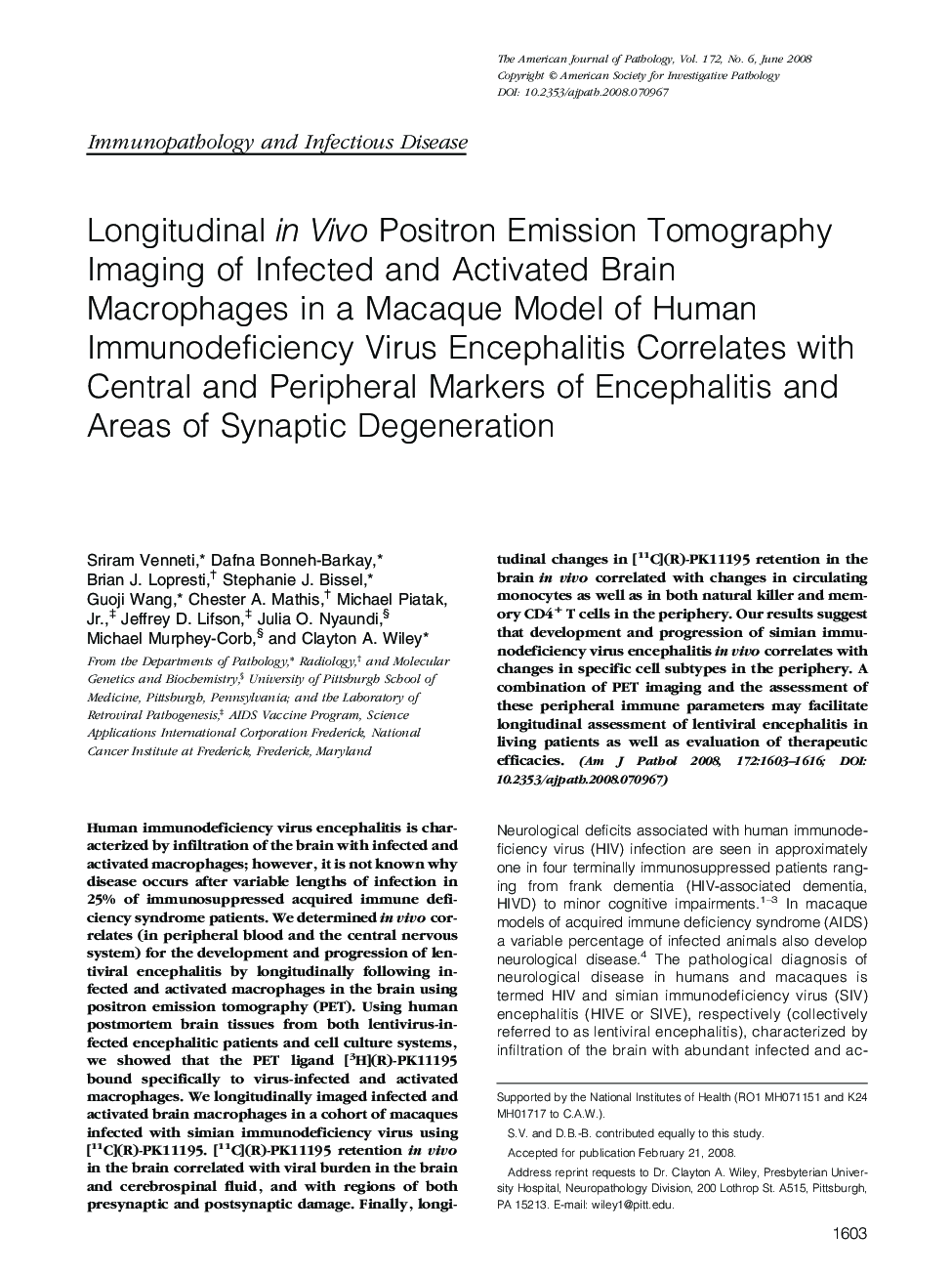
Human immunodeficiency virus encephalitis is characterized by infiltration of the brain with infected and activated macrophages; however, it is not known why disease occurs after variable lengths of infection in 25% of immunosuppressed acquired immune deficiency syndrome patients. We determined in vivo correlates (in peripheral blood and the central nervous system) for the development and progression of lentiviral encephalitis by longitudinally following infected and activated macrophages in the brain using positron emission tomography (PET). Using human postmortem brain tissues from both lentivirus-infected encephalitic patients and cell culture systems, we showed that the PET ligand [3H](R)-PK11195 bound specifically to virus-infected and activated macrophages. We longitudinally imaged infected and activated brain macrophages in a cohort of macaques infected with simian immunodeficiency virus using [11C](R)-PK11195. [11C](R)-PK11195 retention in vivo in the brain correlated with viral burden in the brain and cerebrospinal fluid, and with regions of both presynaptic and postsynaptic damage. Finally, longitudinal changes in [11C](R)-PK11195 retention in the brain in vivo correlated with changes in circulating monocytes as well as in both natural killer and memory CD4+ T cells in the periphery. Our results suggest that development and progression of simian immunodeficiency virus encephalitis in vivo correlates with changes in specific cell subtypes in the periphery. A combination of PET imaging and the assessment of these peripheral immune parameters may facilitate longitudinal assessment of lentiviral encephalitis in living patients as well as evaluation of therapeutic efficacies.
ناشر
Database: Elsevier - ScienceDirect (ساینس دایرکت)
Journal: The American Journal of Pathology - Volume 172, Issue 6, June 2008, Pages 1603-1616
Journal: The American Journal of Pathology - Volume 172, Issue 6, June 2008, Pages 1603-1616
نویسندگان
Sriram Venneti, Dafna Bonneh-Barkay, Brian J. Lopresti, Stephanie J. Bissel, Guoji Wang, Chester A. Mathis, Michael Jr., Jeffrey D. Lifson, Julia O. Nyaundi, Michael Murphey-Corb, Clayton A. Wiley,