| کد مقاله | کد نشریه | سال انتشار | مقاله انگلیسی | نسخه تمام متن |
|---|---|---|---|---|
| 9311347 | 1250202 | 2005 | 20 صفحه PDF | دانلود رایگان |
عنوان انگلیسی مقاله ISI
Athérosclérose et reins
دانلود مقاله + سفارش ترجمه
دانلود مقاله ISI انگلیسی
رایگان برای ایرانیان
کلمات کلیدی
CoronaropathiesAtherosclerosis - آترواسکلروز(تصلب شریان)Cholesterol crystal embolism - آمبولی بلور کلسترولangioplasty - آنژیوپلاستیAngioplastie - آنژیوپلاستیAngiography - آنژیوگرافیAngiographie - آنژیوگرافیStatins - استاتین Statines - استاتین هاrenal ischemia - ایسکمی کلیویCoronary disease - بیماری های عروق کرونریAthérosclérose - تصلب شریانRenal artery stenosis - تنگی شریان های کلیویChirurgie vasculaire - جراحی عروقVascular surgery - جراحی عروقCardiovascular death - مرگ قلب و عروقmortalité cardiovasculaire - مرگ و میر قلب و عروقInsuffisance rénale chronique - نارسایی مزمن کلیهchronic renal insufficiency - نارسایی مزمن کلیویAtheromatous plaque - پلاک آتروماتوز
موضوعات مرتبط
علوم پزشکی و سلامت
پزشکی و دندانپزشکی
بیماریهای کلیوی
پیش نمایش صفحه اول مقاله

چکیده انگلیسی
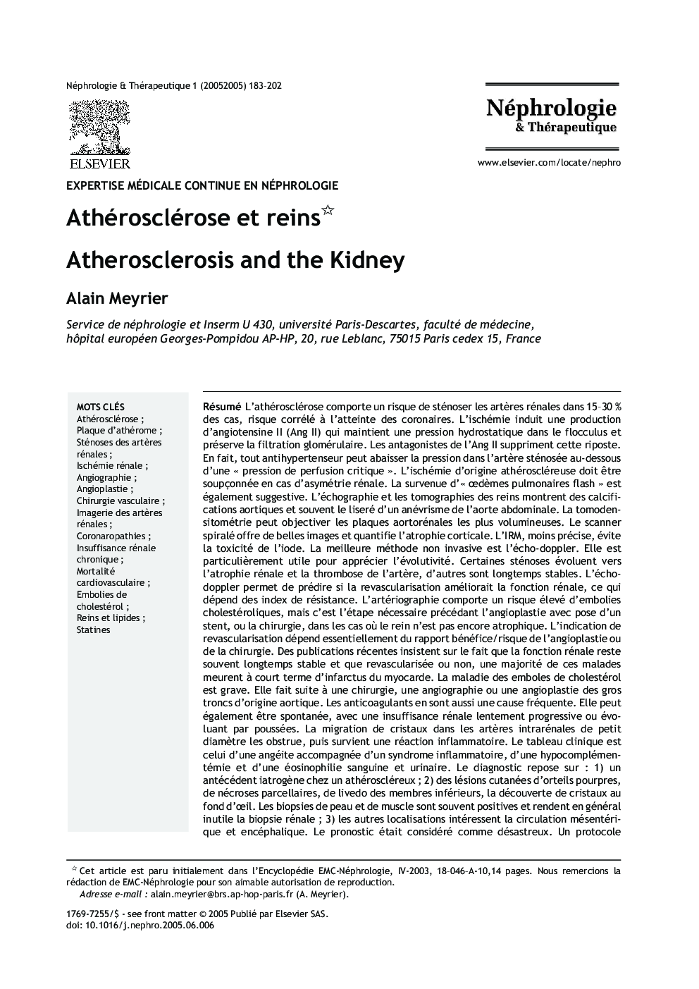
Diffuse atherosclerosis entails a 15-30% risk of plaques on renal arteries (ARAS), with a correlation with coronary atherosclerosis. Ischemia induces generation of angiotensin II (Ang II) that maintains sufficient hydrostatic pressure within the tuft to preserve the GFR. Ang II inhibition suppresses this protective mechanism. In fact, any antihypertensive drug may lead to reaching a "critical perfusion pressure". ARAS should be suspected in case of renal asymmetry. It should also be envisaged in case of "flash pulmonary edemas". Ultrasonography and renal tomography show aortic calcifications and often the outline of an abdominal aortic aneurysm. Tomodensitometry may detect large aorto-renal plaques. Spiral scanner tomography represents a progress, in terms of renal artery imaging and of renal cortical atrophy. Magnetic resonance imaging is less accurate but avoids iodine toxicity. The best noninvasive method is pulsed echo-doppler. It is particularly useful for evaluating stenoses progression. Some stenoses progress to renal atrophy and renal artery thrombosis, whereas others follow a stable course. Pulsed Doppler helps predict whether revascularization will improve renal function, according to the resistance index. Renal arteriography entails a high risk of cholesterol crystal embolism. However, it is the obligatory first step for angioplasty and stent positioning, indicated when the kidney is not atrophic. The indication for revascularization essentially depends on evaluation of the benefits vs risks of angioplasty or surgery. Some publications underscore the frequent stability of renal function and the fact that, revascularized or not, most patients will shortly die of myocardial infarction. Renal cholesterol crystal embolism (CCE) is a severe condition, which occurs when large arteries undergo surgery, aortography or interventional radiology. Anticoagulants are a frequent cause of CCE. CCE may also occur spontaneously, resulting in slowly progressive renal insufficiency. Migration of crystals in small caliber intrarenal arteries induces obstruction, followed by an inflammatory reaction. The clinical picture resembles angiitis, with laboratory evidence of inflammation along with high eosinophil counts and hypocomplementemia. Diagnosis rests on: 1) a iatrogenic event in a patient with an atherosclerotic background; 2) examination of the skin disclosing purple toes, small necrotic lesions and livedo of the lower limbs. Crystals may also be found by funduscopy. Skin or muscle biopsy are contributive in showing crystals and help avoid renal biopsy; 3) other localizations involve the mesenteric circulation and the central nervous system. Until recently, the prognosis was considered disastrous. However, a recently published treatment schedule proved efficient in reducing mortality. A last issue regarding the relationships between atherosclerosis and the kidney deserves mention. In an autopsy-based study it was shown that atherosclerosis per se is accompanied by an increase in the glomerular surface area along with a greater proportion of obsolescent glomeruli by comparison with matched controls. Finally, it should be recalled that atherogenic hyperlipidemia usually aggravates the course of any renal disease, including ARAS. Treatment with statins is indicated in all forms of atherosclerotic renal disease.
ناشر
Database: Elsevier - ScienceDirect (ساینس دایرکت)
Journal: Néphrologie & Thérapeutique - Volume 1, Issue 3, July 2005, Pages 183-202
Journal: Néphrologie & Thérapeutique - Volume 1, Issue 3, July 2005, Pages 183-202
نویسندگان
Alain Meyrier,