| کد مقاله | کد نشریه | سال انتشار | مقاله انگلیسی | نسخه تمام متن |
|---|---|---|---|---|
| 10254943 | 161478 | 2015 | 7 صفحه PDF | دانلود رایگان |
عنوان انگلیسی مقاله ISI
Comparative analysis of Napsin A, alpha-methylacyl-coenzyme A racemase (AMACR, P504S), and hepatocyte nuclear factor 1 beta as diagnostic markers of ovarian clear cell carcinoma: an immunohistochemical study of 279 ovarian tumours
دانلود مقاله + سفارش ترجمه
دانلود مقاله ISI انگلیسی
رایگان برای ایرانیان
موضوعات مرتبط
علوم پزشکی و سلامت
پزشکی و دندانپزشکی
پزشکی قانونی
پیش نمایش صفحه اول مقاله

چکیده انگلیسی
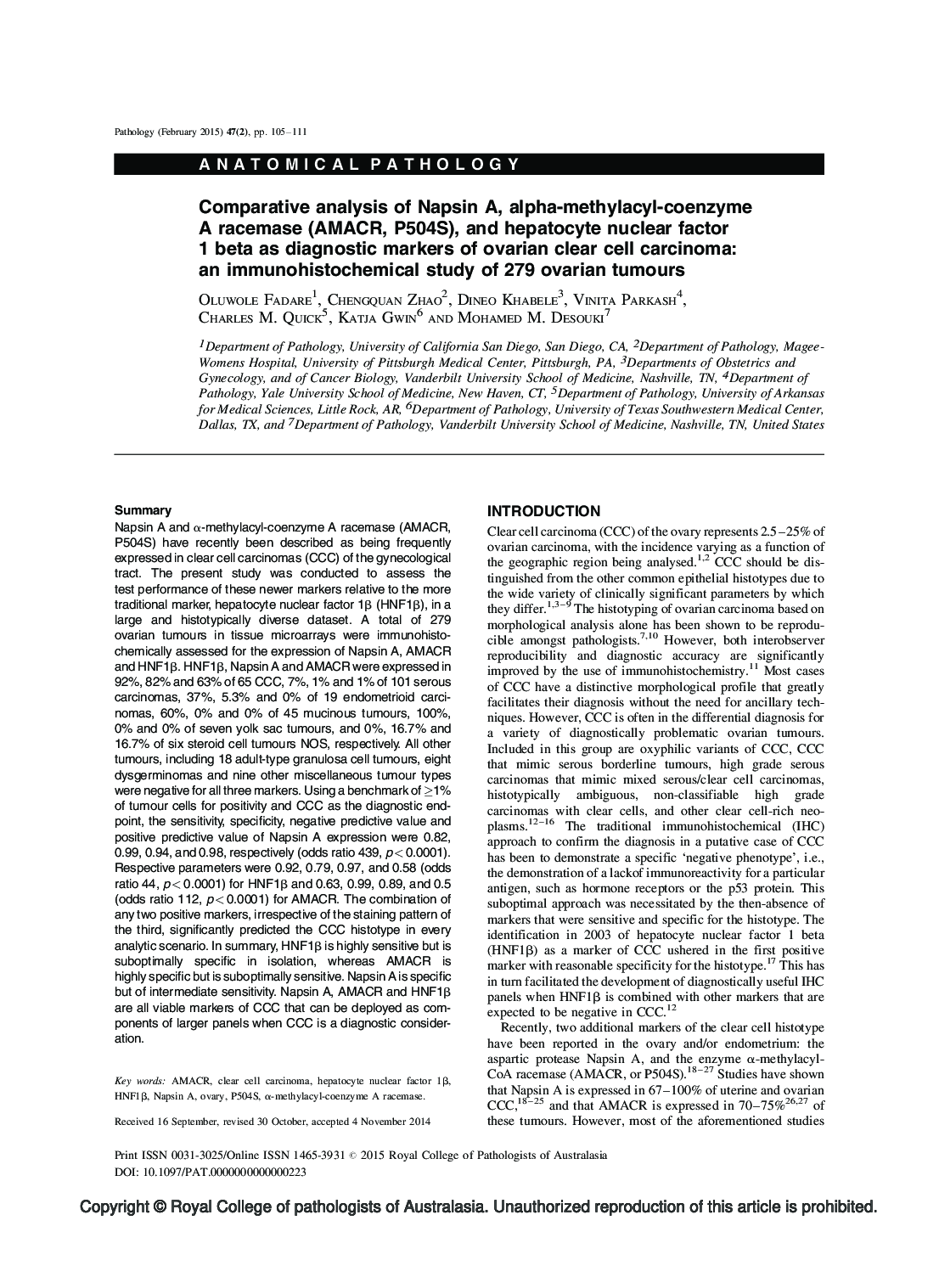
Napsin A and α-methylacyl-coenzyme A racemase (AMACR, P504S) have recently been described as being frequently expressed in clear cell carcinomas (CCC) of the gynecological tract. The present study was conducted to assess the test performance of these newer markers relative to the more traditional marker, hepatocyte nuclear factor 1β (HNF1β), in a large and histotypically diverse dataset. A total of 279 ovarian tumours in tissue microarrays were immunohisto-chemically assessed for the expression of Napsin A, AMACR and HNF1β. HNF1β, Napsin A and AMACR were expressed in 92%, 82% and 63% of 65 CCC, 7%, 1% and 1% of 101 serous carcinomas, 37%, 5.3% and 0% of 19 endometrioid carcinomas, 60%, 0% and 0% of 45 mucinous tumours, 100%, 0% and 0% of seven yolk sac tumours, and 0%, 16.7% and 16.7% of six steroid cell tumours NOS, respectively. All other tumours, including 18 adult-type granulosa cell tumours, eight dysgerminomas and nine other miscellaneous tumour types were negative for all three markers. Using a benchmark of â¥1% of tumour cells for positivity and CCC as the diagnostic end-point, the sensitivity, specificity, negative predictive value and positive predictive value of Napsin A expression were 0.82, 0.99, 0.94, and 0.98, respectively (odds ratio 439, p< 0.0001). Respective parameters were 0.92, 0.79, 0.97, and 0.58 (odds ratio 44, p < 0.0001) for HNF1β and 0.63, 0.99, 0.89, and 0.5 (odds ratio 112, p < 0.0001) for AMACR. The combination of any two positive markers, irrespective of the staining pattern of the third, significantly predicted the CCC histotype in every analytic scenario. In summary, HNF1 β is highly sensitive but is suboptimally specific in isolation, whereas AMACR is highly specific but is suboptimally sensitive. Napsin A is specific but of intermediate sensitivity. Napsin A, AMACR and HNF1β are all viable markers of CCC that can be deployed as components of larger panels when CCC is a diagnostic consideration.
ناشر
Database: Elsevier - ScienceDirect (ساینس دایرکت)
Journal: Pathology - Volume 47, Issue 2, February 2015, Pages 105-111
Journal: Pathology - Volume 47, Issue 2, February 2015, Pages 105-111
نویسندگان
Oluwole Fadare, Chengquan Zhao, Dineo Khabele, Vinita Parkash, Charles M. Quick, Katja Gwin, Mohamed M. Desouki,