| کد مقاله | کد نشریه | سال انتشار | مقاله انگلیسی | نسخه تمام متن |
|---|---|---|---|---|
| 10691647 | 1019602 | 2014 | 13 صفحه PDF | دانلود رایگان |
عنوان انگلیسی مقاله ISI
Early Detection of Liver Fibrosis in Rats Using 3-D Ultrasound Nakagami Imaging: A Feasibility Evaluation
دانلود مقاله + سفارش ترجمه
دانلود مقاله ISI انگلیسی
رایگان برای ایرانیان
کلمات کلیدی
موضوعات مرتبط
مهندسی و علوم پایه
فیزیک و نجوم
آکوستیک و فرا صوت
پیش نمایش صفحه اول مقاله

چکیده انگلیسی
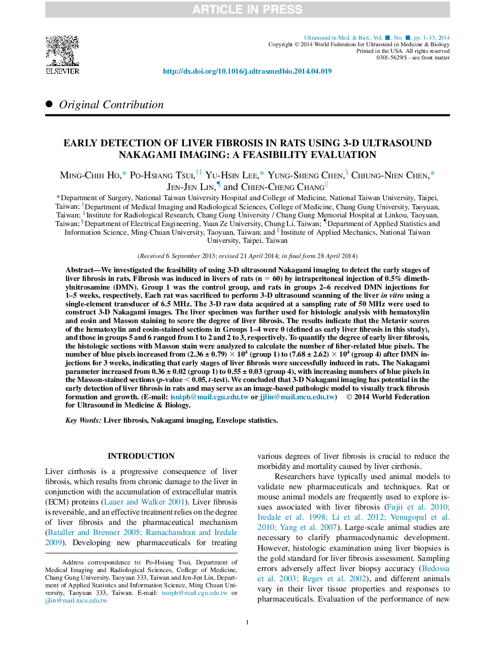
We investigated the feasibility of using 3-D ultrasound Nakagami imaging to detect the early stages of liver fibrosis in rats. Fibrosis was induced in livers of rats (n = 60) by intraperitoneal injection of 0.5% dimethylnitrosamine (DMN). Group 1 was the control group, and rats in groups 2-6 received DMN injections for 1-5 weeks, respectively. Each rat was sacrificed to perform 3-D ultrasound scanning of the liver in vitro using a single-element transducer of 6.5 MHz. The 3-D raw data acquired at a sampling rate of 50 MHz were used to construct 3-D Nakagami images. The liver specimen was further used for histologic analysis with hematoxylin and eosin and Masson staining to score the degree of liver fibrosis. The results indicate that the Metavir scores of the hematoxylin and eosin-stained sections in Groups 1-4 were 0 (defined as early liver fibrosis in this study), and those in groups 5 and 6 ranged from 1 to 2 and 2 to 3, respectively. To quantify the degree of early liver fibrosis, the histologic sections with Masson stain were analyzed to calculate the number of fiber-related blue pixels. The number of blue pixels increased from (2.36 ± 0.79) Ã 104 (group 1) to (7.68 ± 2.62) Ã 104 (group 4) after DMN injections for 3 weeks, indicating that early stages of liver fibrosis were successfully induced in rats. The Nakagami parameter increased from 0.36 ± 0.02 (group 1) to 0.55 ± 0.03 (group 4), with increasing numbers of blue pixels in the Masson-stained sections (p-value < 0.05, t-test). We concluded that 3-D Nakagami imaging has potential in the early detection of liver fibrosis in rats and may serve as an image-based pathologic model to visually track fibrosis formation and growth.
ناشر
Database: Elsevier - ScienceDirect (ساینس دایرکت)
Journal: Ultrasound in Medicine & Biology - Volume 40, Issue 9, September 2014, Pages 2272-2284
Journal: Ultrasound in Medicine & Biology - Volume 40, Issue 9, September 2014, Pages 2272-2284
نویسندگان
Ming-Chih Ho, Po-Hsiang Tsui, Yu-Hsin Lee, Yung-Sheng Chen, Chiung-Nien Chen, Jen-Jen Lin, Chien-Cheng Chang,