| کد مقاله | کد نشریه | سال انتشار | مقاله انگلیسی | نسخه تمام متن |
|---|---|---|---|---|
| 1760431 | 1019590 | 2015 | 8 صفحه PDF | دانلود رایگان |
عنوان انگلیسی مقاله ISI
High-Frequency Ultrasound as an Option for Scanning of Prepared Teeth: An in Vitro Study
دانلود مقاله + سفارش ترجمه
دانلود مقاله ISI انگلیسی
رایگان برای ایرانیان
موضوعات مرتبط
مهندسی و علوم پایه
فیزیک و نجوم
آکوستیک و فرا صوت
پیش نمایش صفحه اول مقاله

چکیده انگلیسی
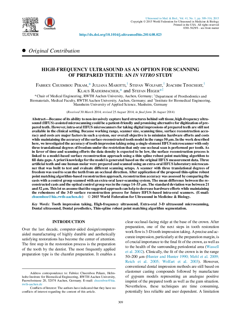
Because of its ability to non-invasively capture hard structures behind soft tissue, high-frequency ultrasound (HFUS)-assisted microscanning could be a patient-friendly and promising alternative for digitization of prepared teeth. However, intra-oral HFUS microscanners for taking digital impressions of prepared teeth are still not available in the clinical setting. Because working range, scanner size, scanning time, surface reconstruction accuracy and costs are major factors in such a system, our overall objective is to minimize hardware efforts and costs while maintaining the accuracy of the surface-reconstructed tooth model in the range 50 μm. In the work described here, we investigated the accuracy of tooth impression taking using a single-element HFUS microscanner with only three translational degrees of freedom under the restriction that only one occlusal scan is performed per tooth. As in favor of time and scanning efforts the data density is expected to be low, the surface reconstruction process is linked to a model-based surface reconstruction approach using a thin spline robust point matching algorithm to fill data gaps. A priori knowledge for the model is generated based on the original HFUS measurement data. Three artificial teeth and one human molar were prepared and scanned using an extra-oral HFUS laboratory microscanner that was built to test and evaluate different scanning setups. A scanner with three translational degrees of freedom was used to scan the teeth from an occlusal direction. After application of the proposed thin-spline robust point matching algorithm-based reconstruction approach, reconstruction accuracy was assessed by comparing the casts with a control group scanned with an extra-oral laser-scanning system. The mean difference between the reconstructed casts and the optical control group was in the range 14-53 μm. The standard deviation was between 21 and 52 μm. This let us assume that the suggested approach can help to decrease hardware efforts while maintaining the robustness of the 3-D surface reconstruction process for future HFUS-based intra-oral scanners.
ناشر
Database: Elsevier - ScienceDirect (ساینس دایرکت)
Journal: Ultrasound in Medicine & Biology - Volume 41, Issue 1, January 2015, Pages 309-316
Journal: Ultrasound in Medicine & Biology - Volume 41, Issue 1, January 2015, Pages 309-316
نویسندگان
Fabrice Chuembou Pekam, Juliana Marotti, Stefan Wolfart, Joachim Tinschert, Klaus Radermacher, Stefan Heger,