| کد مقاله | کد نشریه | سال انتشار | مقاله انگلیسی | نسخه تمام متن |
|---|---|---|---|---|
| 1761049 | 1019631 | 2011 | 9 صفحه PDF | دانلود رایگان |
عنوان انگلیسی مقاله ISI
Comparison of Scanning Acoustic Microscopy and Histology Images in Characterizing Surface Irregularities Among Engineered Human Oral Mucosal Tissues
دانلود مقاله + سفارش ترجمه
دانلود مقاله ISI انگلیسی
رایگان برای ایرانیان
موضوعات مرتبط
مهندسی و علوم پایه
فیزیک و نجوم
آکوستیک و فرا صوت
پیش نمایش صفحه اول مقاله

چکیده انگلیسی
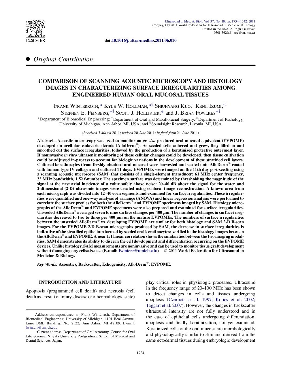
Acoustic microscopy was used to monitor an ex vivo produced oral mucosal equivalent (EVPOME) developed on acellular cadaveric dermis (AlloDerm®). As seeded cells adhered and grew, they filled in and smoothed out the surface irregularities, followed by the production of a keratinized protective outermost layer. If noninvasive in vitro ultrasonic monitoring of these cellular changes could be developed, then tissue cultivation could be adjusted in-process to account for biologic variations in the development of these stratified cell layers. Cultured keratinocytes (from freshly obtained oral mucosa) were harvested and seeded onto AlloDerm® coated with human type IV collagen and cultured 11 days. EVPOMEs were imaged on the 11th day post-seeding using a scanning acoustic microscope (SAM) that consists of a single-element transducer: 61 MHz center frequency, 32 MHz bandwidth, 1.52 f-number. The specimen surface was determined by thresholding the magnitude of the signal at the first axial incidence of a value safely above noise: 20-40 dB above the signal for the water and 2-dimensional (2-D) ultrasonic images were created using confocal image reconstruction. A known area from each micrograph was divided into 12-40 even segments and examined for surface irregularities. These irregularities were quantified and one-way analysis of variance (ANOVA) and linear regression analysis were performed to correlate the surface profiles for both the AlloDerm® and EVPOME specimens imaged by SAM. Histology micrographs of the AlloDerm® and EVPOME specimens were also prepared and examined for surface irregularities. Unseeded AlloDerm® averaged seven to nine surface changes per 400 μm. The number of changes in surface irregularities decreased to two to three per 400 μm on the mature EVPOMEs. The numbers of surface irregularities between the unseeded AlloDerm® vs. developing EVPOME are similar for both histology and SAM 2-D B-scan images. For the EVPOME 2-D B-scan micrographs produced by SAM, the decrease in surface irregularities is indicative of the stratified epithelium formed by seeded oral keratinocytes; verified in the histology images between the AlloDerm® and EVPOME. A near 1:1 linear correlation shows the similarities between the two imaging modalities. SAM demonstrates its ability to discern the cell development and differentiation occurring on the EVPOME devices. Unlike histology, SAM measurements are noninvasive and can be used to monitor tissue graft development without damaging any cells/tissues.
ناشر
Database: Elsevier - ScienceDirect (ساینس دایرکت)
Journal: Ultrasound in Medicine & Biology - Volume 37, Issue 10, October 2011, Pages 1734-1742
Journal: Ultrasound in Medicine & Biology - Volume 37, Issue 10, October 2011, Pages 1734-1742
نویسندگان
Frank Winterroth, Kyle W. Hollman, Shiuhyang Kuo, Kenji Izumi, Stephen E. Feinberg, Scott J. Hollister, J. Brian Fowlkes,