| کد مقاله | کد نشریه | سال انتشار | مقاله انگلیسی | نسخه تمام متن |
|---|---|---|---|---|
| 1761470 | 1019649 | 2012 | 11 صفحه PDF | دانلود رایگان |
عنوان انگلیسی مقاله ISI
Speckle Noise Reduction in Ultrasound Biomedical B-Scan Images Using Discrete Topological Derivative
دانلود مقاله + سفارش ترجمه
دانلود مقاله ISI انگلیسی
رایگان برای ایرانیان
موضوعات مرتبط
مهندسی و علوم پایه
فیزیک و نجوم
آکوستیک و فرا صوت
پیش نمایش صفحه اول مقاله

چکیده انگلیسی
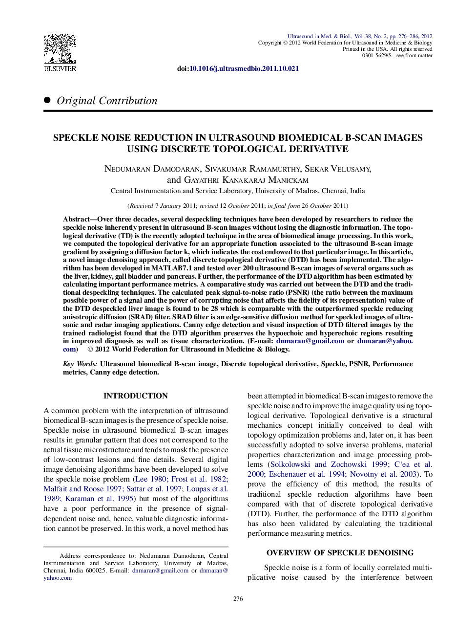
Over three decades, several despeckling techniques have been developed by researchers to reduce the speckle noise inherently present in ultrasound B-scan images without losing the diagnostic information. The topological derivative (TD) is the recently adopted technique in the area of biomedical image processing. In this work, we computed the topological derivative for an appropriate function associated to the ultrasound B-scan image gradient by assigning a diffusion factor k, which indicates the cost endowed to that particular image. In this article, a novel image denoising approach, called discrete topological derivative (DTD) has been implemented. The algorithm has been developed in MATLAB7.1 and tested over 200 ultrasound B-scan images of several organs such as the liver, kidney, gall bladder and pancreas. Further, the performance of the DTD algorithm has been estimated by calculating important performance metrics. A comparative study was carried out between the DTD and the traditional despeckling techniques. The calculated peak signal-to-noise ratio (PSNR) (the ratio between the maximum possible power of a signal and the power of corrupting noise that affects the fidelity of its representation) value of the DTD despeckled liver image is found to be 28 which is comparable with the outperformed speckle reducing anisotropic diffusion (SRAD) filter. SRAD filter is an edge-sensitive diffusion method for speckled images of ultrasonic and radar imaging applications. Canny edge detection and visual inspection of DTD filtered images by the trained radiologist found that the DTD algorithm preserves the hypoechoic and hyperechoic regions resulting in improved diagnosis as well as tissue characterization.
ناشر
Database: Elsevier - ScienceDirect (ساینس دایرکت)
Journal: Ultrasound in Medicine & Biology - Volume 38, Issue 2, February 2012, Pages 276-286
Journal: Ultrasound in Medicine & Biology - Volume 38, Issue 2, February 2012, Pages 276-286
نویسندگان
Nedumaran Damodaran, Sivakumar Ramamurthy, Sekar Velusamy, Gayathri Kanakaraj Manickam,