| کد مقاله | کد نشریه | سال انتشار | مقاله انگلیسی | نسخه تمام متن |
|---|---|---|---|---|
| 1762657 | 1019708 | 2008 | 9 صفحه PDF | دانلود رایگان |
عنوان انگلیسی مقاله ISI
Hemorrhage Detection During Focused-Ultrasound Induced Blood-Brain-Barrier Opening by Using Susceptibility-Weighted Magnetic Resonance Imaging
دانلود مقاله + سفارش ترجمه
دانلود مقاله ISI انگلیسی
رایگان برای ایرانیان
کلمات کلیدی
موضوعات مرتبط
مهندسی و علوم پایه
فیزیک و نجوم
آکوستیک و فرا صوت
پیش نمایش صفحه اول مقاله

چکیده انگلیسی
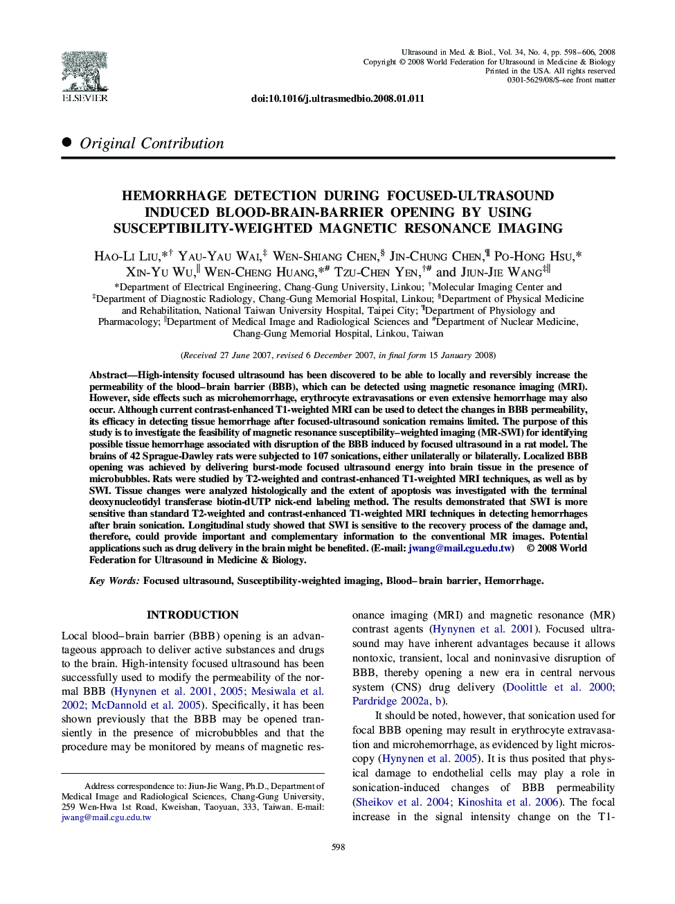
High-intensity focused ultrasound has been discovered to be able to locally and reversibly increase the permeability of the blood-brain barrier (BBB), which can be detected using magnetic resonance imaging (MRI). However, side effects such as microhemorrhage, erythrocyte extravasations or even extensive hemorrhage may also occur. Although current contrast-enhanced T1-weighted MRI can be used to detect the changes in BBB permeability, its efficacy in detecting tissue hemorrhage after focused-ultrasound sonication remains limited. The purpose of this study is to investigate the feasibility of magnetic resonance susceptibility-weighted imaging (MR-SWI) for identifying possible tissue hemorrhage associated with disruption of the BBB induced by focused ultrasound in a rat model. The brains of 42 Sprague-Dawley rats were subjected to 107 sonications, either unilaterally or bilaterally. Localized BBB opening was achieved by delivering burst-mode focused ultrasound energy into brain tissue in the presence of microbubbles. Rats were studied by T2-weighted and contrast-enhanced T1-weighted MRI techniques, as well as by SWI. Tissue changes were analyzed histologically and the extent of apoptosis was investigated with the terminal deoxynucleotidyl transferase biotin-dUTP nick-end labeling method. The results demonstrated that SWI is more sensitive than standard T2-weighted and contrast-enhanced T1-weighted MRI techniques in detecting hemorrhages after brain sonication. Longitudinal study showed that SWI is sensitive to the recovery process of the damage and, therefore, could provide important and complementary information to the conventional MR images. Potential applications such as drug delivery in the brain might be benefited. (E-mail: jwang@mail.cgu.edu.tw)
ناشر
Database: Elsevier - ScienceDirect (ساینس دایرکت)
Journal: Ultrasound in Medicine & Biology - Volume 34, Issue 4, April 2008, Pages 598-606
Journal: Ultrasound in Medicine & Biology - Volume 34, Issue 4, April 2008, Pages 598-606
نویسندگان
Hao-Li Liu, Yau-Yau Wai, Wen-Shiang Chen, Jin-Chung Chen, Po-Hong Hsu, Xin-Yu Wu, Wen-Cheng Huang, Tzu-Chen Yen, Jiun-Jie Wang,