| کد مقاله | کد نشریه | سال انتشار | مقاله انگلیسی | نسخه تمام متن |
|---|---|---|---|---|
| 2768950 | 1151287 | 2010 | 15 صفحه PDF | دانلود رایگان |
عنوان انگلیسی مقاله ISI
La punción guiada con ultrasonidos aplicada a una unidad de dolor crónico
دانلود مقاله + سفارش ترجمه
دانلود مقاله ISI انگلیسی
رایگان برای ایرانیان
کلمات کلیدی
موضوعات مرتبط
علوم پزشکی و سلامت
پزشکی و دندانپزشکی
بیهوشی و پزشکی درد
پیش نمایش صفحه اول مقاله

چکیده انگلیسی
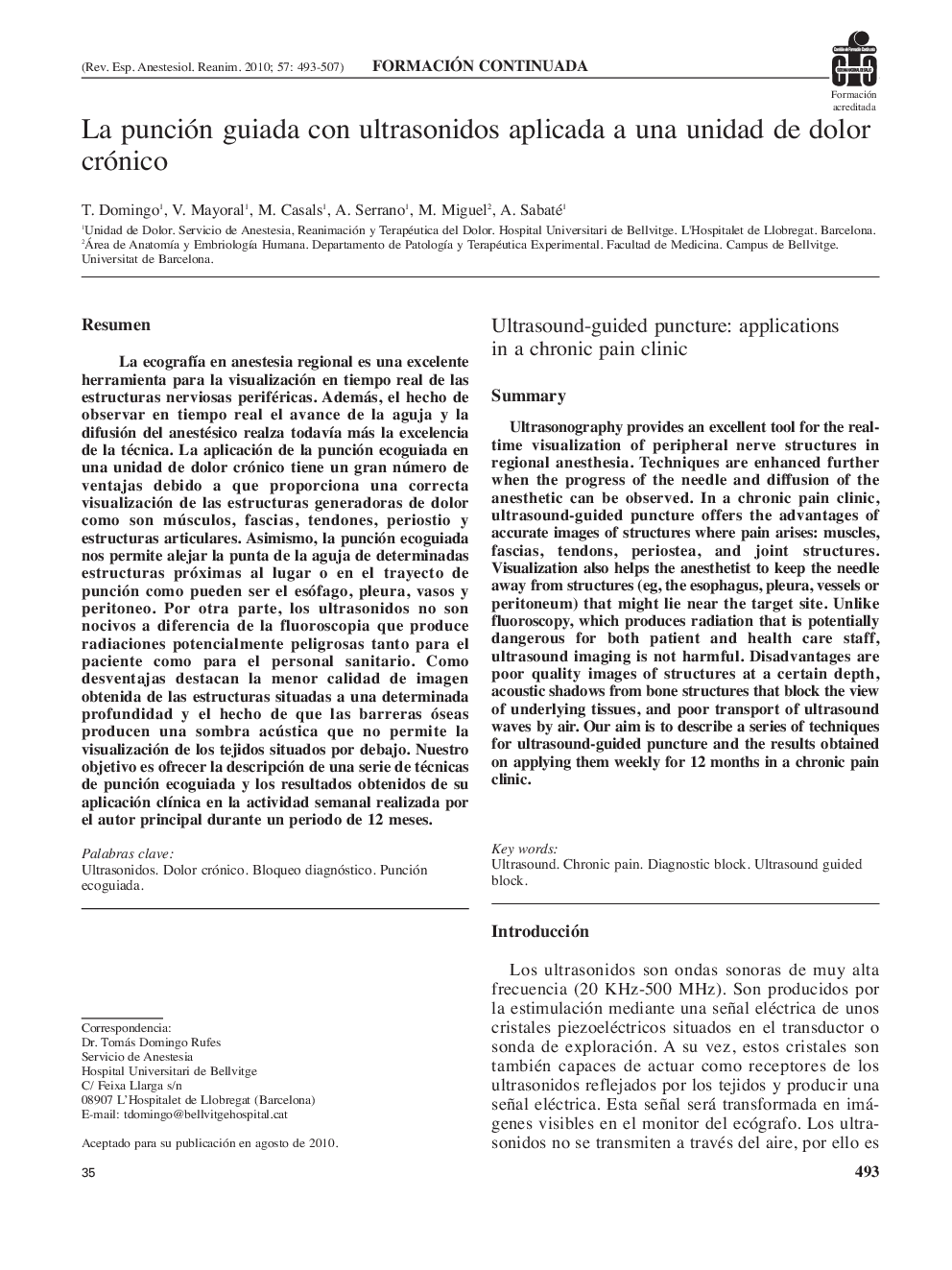
Ultrasonography provides an excellent tool for the real-time visualization of peripheral nerve structures in regional anesthesia. Techniques are enhanced further when the progress of the needle and diffusion of the anesthetic can be observed. In a chronic pain clinic, ultrasound-guided puncture offers the advantages of accurate images of structures where pain arises: muscles, fascias, tendons, periostea, and joint structures. Visualization also helps the anesthetist to keep the needle away from structures (eg, the esophagus, pleura, vessels or peritoneum) that might lie near the target site. Unlike fluoroscopy, which produces radiation that is potentially dangerous for both patient and health care staff, ultrasound imaging is not harmful. Disadvantages are poor quality images of structures at a certain depth, acoustic shadows from bone structures that block the view of underlying tissues, and poor transport of ultrasound waves by air. Our aim is to describe a series of techniques for ultrasound-guided puncture and the results obtained on applying them weekly for 12 months in a chronic pain clinic.
ناشر
Database: Elsevier - ScienceDirect (ساینس دایرکت)
Journal: Revista Española de AnestesiologÃa y Reanimación - Volume 57, Issue 8, 2010, Pages 493-507
Journal: Revista Española de AnestesiologÃa y Reanimación - Volume 57, Issue 8, 2010, Pages 493-507
نویسندگان
T. Domingo, V. Mayoral, M. Casals, A. Serrano, M. Miguel, A. Sabaté,