| کد مقاله | کد نشریه | سال انتشار | مقاله انگلیسی | نسخه تمام متن |
|---|---|---|---|---|
| 6276739 | 1295742 | 2010 | 13 صفحه PDF | دانلود رایگان |
عنوان انگلیسی مقاله ISI
Proline-rich synapse-associated protein-1 and 2 (ProSAP1/Shank2 and ProSAP2/Shank3)-scaffolding proteins are also present in postsynaptic specializations of the peripheral nervous system
دانلود مقاله + سفارش ترجمه
دانلود مقاله ISI انگلیسی
رایگان برای ایرانیان
کلمات کلیدی
α-bungarotoxinmembrane-associated guanylate kinasesSAPAPIntraganglionic laminar endingIntraganglionic laminar endingssynaptic cleftSIBVGLUTVAChTN-methyl-d-aspartic acidENSGKAPmGluRDABNADPH-dNMDANMJGFAPPSDSCGnAChRsuperior cervical ganglion - ganglion برتر گردن رحمMAGUK - آنهاNeuromuscular junction - اتصال عصبی عضلانیMotor endplate - انداختن موتورimmunoreactive - ایمنی فعالImmunoreactivity - ایمنی فعالpostsynaptic density - تراکم Postinaptictyrosine hydroxylase - تیروزین هیدروکسیلازvesicular glutamate transporter - حمل و نقل گلوتامات vesicularvesicular acetylcholine transporter - حمل کننده استیل کولین vesicularAutonomic nervous system - دستگاه عصبی خودمختار یا خودگردان یا اتونومdiaminobenzidine - دیامینو بنزیدینANS - سالenteric nervous system - سیستم عصبی روده ایperipheral nervous system - سیستم عصبی پیرامونیSynaptophysin - سیناپتوفیزینSyn - سینتActive zone - منطقه فعالneurofilament - نوروفیلامنت-ir - وGlial fibrillary acidic protein - پروتئین اسیدی فیبریلاسیون گلایالPNS - کارمندان دولتSynaptic vesicles - کیسه های سیناپتیکnicotinic acetylcholine receptor - گیرنده استیلکولین نیکوتینMetabotropic glutamate receptor - گیرنده گلوتامات متابوتروپیک
موضوعات مرتبط
علوم زیستی و بیوفناوری
علم عصب شناسی
علوم اعصاب (عمومی)
پیش نمایش صفحه اول مقاله

چکیده انگلیسی
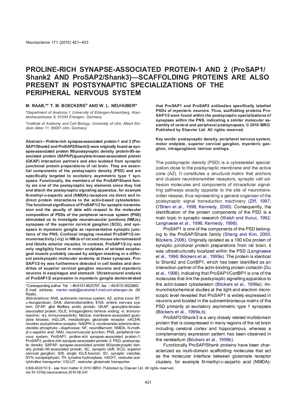
Proline-rich synapse-associated protein-1 and 2 (ProSAP1/Shank2 and ProSAP2/Shank3) were originally found as synapse-associated protein 90/postsynaptic density protein-95-associated protein (SAPAP)/guanylate-kinase-associated protein (GKAP) interaction partners and also isolated from synaptic junctional protein preparations of rat brain. They are essential components of the postsynaptic density (PSD) and are specifically targeted to excitatory asymmetric type 1 synapses. Functionally, the members of the ProSAP/Shank family are one of the postsynaptic key elements since they link and attach the postsynaptic signaling apparatus, for example N-methyl-d-aspartic acid (NMDA)-receptors via direct and indirect protein interactions to the actin-based cytoskeleton. The functional significance of ProSAP1/2 for synaptic transmission and the paucity of data with respect to the molecular composition of PSDs of the peripheral nervous system (PNS) stimulated us to investigate neuromuscular junctions (NMJs), synapses of the superior cervical ganglion (SCG), and synapses in myenteric ganglia as representative synaptic junctions of the PNS. Confocal imaging revealed ProSAP1/2-immunoreactivity (-iry) in NMJs of rat and mouse sternomastoid and tibialis anterior muscles. In contrast, ProSAP1/2-iry was only negligibly found in motor endplates of striated esophageal muscle probably caused by antigen masking or a different postsynaptic molecular anatomy at these synapses. ProSAP1/2-iry was furthermore detected in cell bodies and dendrites of superior cervical ganglion neurons and myenteric neurons in esophagus and stomach. Ultrastructural analysis of ProSAP1/2 expression in myenteric ganglia demonstrated that ProSAP1 and ProSAP2 antibodies specifically labelled PSDs of myenteric neurons. Thus, scaffolding proteins ProSAP1/2 were found within the postsynaptic specializations of synapses within the PNS, indicating a similar molecular assembly of central and peripheral postsynapses.
ناشر
Database: Elsevier - ScienceDirect (ساینس دایرکت)
Journal: Neuroscience - Volume 171, Issue 2, 1 December 2010, Pages 421-433
Journal: Neuroscience - Volume 171, Issue 2, 1 December 2010, Pages 421-433
نویسندگان
M. Raab, T.M. Boeckers, W.L. Neuhuber,